DOI:
10.37988/1811-153X_2026_1_142Clinical and radiological results of coronectomy of the third molars adjacent to the mandibular canal: case series
Downloads
Abstract
A method of performing a coronectomy is presented as an incomplete extraction of the third lower molars. The described technique is based on the prevention of damage to the structures of the mandibular canal in cases of their close interposition with the tooth, and also helps to reduce the time of intervention and its invasiveness. In 3 clinical cases, the features of the postoperative period and the X-ray picture of this surgical treatment method were demonstrated.Results.
Of the 22 coronectomy operations performed on the lower third molars, none showed signs of damage to the inferior alveolar nerve. There were no signs of wound dehiscence, pulpitis, or the development of an infectious and inflammatory process in the postoperative area in all patients associated with coronectomy surgery. In one case (5%), repeated intervention was performed — extraction of the roots due to their eruption into the oral cavity.
Key words:
third molar removal, cone-beam computed tomography, nerve paresthesia, coronectomyFor Citation
Introduction
Lower third molars extraction is one of the most common dental surgical procedures [1]. In addition to surgical indications for extraction such as periodontitis, pericoronitis, and periostitis, intact and asymptomatic third molars are now often extracted in preparation for orthodontic treatment and to prevent malocclusion. Also, if the crown of the third molar is mesially inclined, there is a risk of damage to the adjacent tooth, namely the formation of caries of the crown or root of the tooth or pathological resorption of tooth at the contact point [2, 3].
A combination of anatomical factors, namely the smallest distance among all teeth from the edge of the alveolar arch to the mandibular canal in the area of third molar [4], as well as the most frequent its retention and dystopia [5], leads to the intimate interrelation of the third molar with the inferior alveolar nerve (IAN). Extraction of such teeth is associated with an additional risk of complications in the form of IAN trauma, accompanied by paresthesia. According to the literature, this complication occurs in 0.35–8.4% of patients [6, 7] and on average in 2.9% of cases [8]. The duration of paresthesia can vary significantly, from several days to several months.
Nerve damage is divided into three main categories: neuropraxia, axonotmesis, and neurotmesis. Neuropraxia is the mildest form and is often the result of blunt trauma or pressure. With neuropraxia, spontaneous recovery tends to occur quickly. Axonotmesis is often the result of crushing. Recovery can take anywhere from six months to a year, but depending on the nature and severity of the injury, it can lead to irreversible dysfunction. Neurotmesis is a complete rupture of the nerve, and microsurgical repair is required to achieve significant recovery [9].
The area of paresthesia can cover either a small area of skin or oral mucosa corresponding to the area of innervation of the trigeminal nerve, or the entire area, leading to patient discomfort, speech impairment, difficulty eating and drinking, and an increased risk of acute and chronic trauma to the oral mucosa [10]. Although the prevalence of IAN trauma is low among other possible complications of lower third molar extraction surgery, it significantly affects the patient's quality of life, and its treatment is difficult, since there is no clear treatment protocol [11–13] and, accordingly, practicing dental surgeons should avoid the occurrence of this iatrogenic complication.
Given the above, thorough diagnosis of each clinical case of third molar extraction using modern research methods is of paramount importance. It is also important to apply surgical protocols in clinical practice that allow, on the one hand, to solve the clinical task and, on the other hand, to avoid trauma to the IAN.
Domestic and foreign authors note seven signs on an orthopantomogram that may indicate a high risk of damage to the IAN during the extraction of third molar [14, 15]:
- the presence of an apical shadow in the root apex area, darkening of the bone tissue in the root area (due to decreased bone density);
- deviation/tilt of the tooth roots (sharp bend of the root near the mandibular canal);
- narrowing of the tooth roots at the intersection of the mandibular canal;
- double root tips affected by the mandibular canal;
- rupture of the mandibular canal wall (interruption of the white line);
- diversion of the mandibular canal (change in the direction of the canal in the area of the roots of the third molars);
- narrowing of the mandibular canal (decrease in the diameter of the mandibular canal at the points of intersection with the tips of the roots of the third molars).
Currently, the gold standard for diagnosis is cone beam computed tomography (CBCT), which allows visualization of the mandibular nerve and thus provides maximum information about the anatomy and path of the mandibular canal, as well as its relationship to the roots of the mandibular teeth in three-dimensional space [16].
However, thorough radiological diagnosis of each clinical case only helps to assess the risks of complications during tooth extraction in order to warn the patient, but does not allow them to be avoided, especially when the doctor, knowing about the high risks, proceeds with complete extraction of the third molar. In addition, there are many cases when, based on X-ray data on the relative position of the roots of the third molar and the IAN, the patient may be denied outpatient treatment and referred to a maxillofacial surgery hospital, justifying the refusal by the increased complexity of the operation and possible complications associated with trauma to the IAN. However, the protocol for complete tooth extraction in a hospital setting is no different from surgical treatment in an outpatient setting, and such intervention also carries similar risks of complications.
One way to avoid traumatizing the pulp is coronectomy [17]. According to this treatment method, only the crown of the tooth covered with enamel is removed, while the root of the tooth is left in the alveolar bone. The tooth undergoing coronectomy must be vital: intact or with signs of a carious cavity that does not affected the pulp and without any signs of pathological changes in the periodontium [18].
There are very few domestic scientific studies on this technique with long-term clinical results, and awareness among doctors, including those with extensive experience, is low [19]. In the domestic professional sphere, coronectomy is currently classified as a controversial surgical procedure. Questions remain unresolved regarding the surgical protocol for the operation, the need for endodontic treatment of the remaining roots, and the characteristics of the early and late postoperative periods. There is also the fact that the remaining root fragments may be a potential source of infection. In particular, any inflammatory odontogenic processes on the side where coronectomy was previously performed may be clinically associated with the remaining root, even if it is not a source of infection, which complicates diagnosis. Legally, the dentist who performed the incomplete tooth extraction becomes responsible for any possible inflammatory complications in this area, which increases the need for more research.
The aim of the study is to demonstrate the clinical efficacy of coronectomy of the lower third molars of in cases of high risk of trauma to the IAN.
Materials and methods
The authors performed a total of 22 coronectomy operations on lower third molars in 21 adult patients aged 18–42 years with a follow-up period of up to 2.5 years. When planning the extraction of vital lower third molars, all patients showed radiographic signs of tooth root contact with the mandibular canal on CBCT with no signs of pulpitis or periodontitis.
At the point of choosing a treatment strategy, both complete tooth extraction as the main treatment method and coronectomy were discussed with the patients. Patients were given a detailed description of the pros and cons of the proposed treatment options, possible complications, the course of the operation, and the likelihood of a repeat visit in case of coronal root migration, root eruption into the oral cavity, or inflammation in the area of the tooth roots with a previous coronectomy.
When choosing coronectomy as a treatment, patients underwent CBCT examination before and after surgery, after 3–4 months, and then every 6 months. During CBCT analysis, images of sections in the sagittal and frontal planes were obtained. To assess the “eruption” of the third molar roots, the minimum distance from the roots of the tooth that underwent coronectomy to the cementoenamel junction (CEJ) of the adjacent mesial tooth was measured on the sagittal section. In a graphics editor, the boundaries of the IAN and tooth roots were highlighted for better visualization to assess their relative positions.
Follow-up examinations were conducted 7–10 days, 3–4 months, and 6 months after surgery, and then every 6 months thereafter.
Surgical protocol
After antiseptic rinse of the oral cavity with chlorhexidine (0.05%) mouthwashes for 1 minute (Fig. 1A), local anesthesia was performed. Benzocaine gel was applied to the surgical area, and conduction mandibular anesthesia with aspiration test and infiltration anesthesia (4% articaine solution with 1:200,000 adrenaline with a total volume of up to 3.4 ml) along the transitional fold in the area of the third molar from the vestibular side.
In the retromolar region, a horizontal incision was made with a distal vertical incision directed toward the transitional fold and a vestibular intrasulcular incision in the region of the second molar with a vertical relaxing incision in the region of the mesial edge of the crown of the second molar, and a full-thickness flap was raised. Next step pericoronal osteotomy was performed using a straight surgical tip and a carbide bur with external cooling with saline solution to form surgical access and visualize the crown part of the tooth (Fig. 1B). Using an high speed contraangled tip with a highly abrasive diamond bur and external cooling with saline solution, the crown part of the tooth was removed to at least the level of the cementoenamel junction (Fig. 1C).
At this stage of coronectomy, doctors should pay special attention to the distal and distolingual parts of the tooth, as these areas remain enamel the most difficult to access when the tooth is deeply embedded. The preparation margin around the perimeter of the tooth is located at or below the level of the bone tissue to allow for subsequent wound closure and minimize subsequent eruption of the remaining tooth fragment. If possible, the crown of the tooth should be completely separated with a bur, but in cases of high risk of trauma to soft tissues, including the lingual nerve and the inferior alveolar nerve, as well as the adjacent second molar, approximately 1 mm of the tooth crown tissue should be left intact and the crown should be broken off with a slight rotational movement of the elevator without excessive force. After removing the crown of the tooth, no endodontic treatment, root canal preparation, or pulp extirpation of the remaining roots was performed.
After removing the crown of the tooth, the wound was treated with chlorhexidine rinsing and wound hemostasis was achieved by tamponade with a sterile gauze swab. A control intraoral periapical radiograph of the surgical area was taken to check for the presence of enamel areas. After completing the preparation, the pulp and debris residues were washed out with chlorhexidine (0.05%), and curettage of the socket was performed only in the crown part of the socket. Repeated hemostasis of the wound was performed, and the flap was put back in place.
If necessary, vestibular flaps were mobilized to close the wound completely. The flaps were fixed with monofilament non-absorbable suture material using simple knot sutures (Fig. 1D).
After the operation, all patients were prescribed a gentle diet and irrigation of the oral cavity with chlorhexidine (0.05%) three times a day for 7 days, starting the day after the operation. Taking into account the medical history, antibiotic therapy (500 mg of amoxicillin with 125 mg of clavulanic acid) was prescribed every 8 hours for 5 days. For pain, NSAIDs were recommended every 4–6 hours in accordance with the manufacturer's recommendations. Oral care was prescribed in the form of brushing teeth twice a day with a soft toothbrush and toothpaste, avoiding the area of intervention for 7 days.
A second routine checkup for suture removal was performed on day 7–10.
Results
Of the 22 coronectomy operations performed on lower third molars, no signs of damage to the IAN were noted in any case. No signs of wound edge dehiscence, pulpitis, or the development of an infectious-inflammatory process in the postoperative area associated with coronectomy surgery were detected in any of the patients. In one case (4.5%), a repeat intervention was performed — extraction of the roots due to their eruption into the oral cavity. Further observation of the patients included in this study is ongoing.
The capabilities of the technique will be further demonstrated in 3 clinical cases.
Clinical case 1
Patient T., 25 years old, complained of difficult tooth eruption and periodic pain in the right lower segment of jaw.
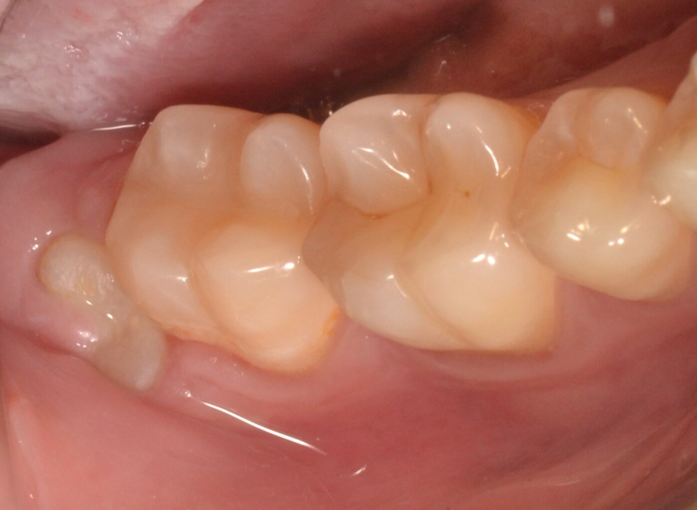
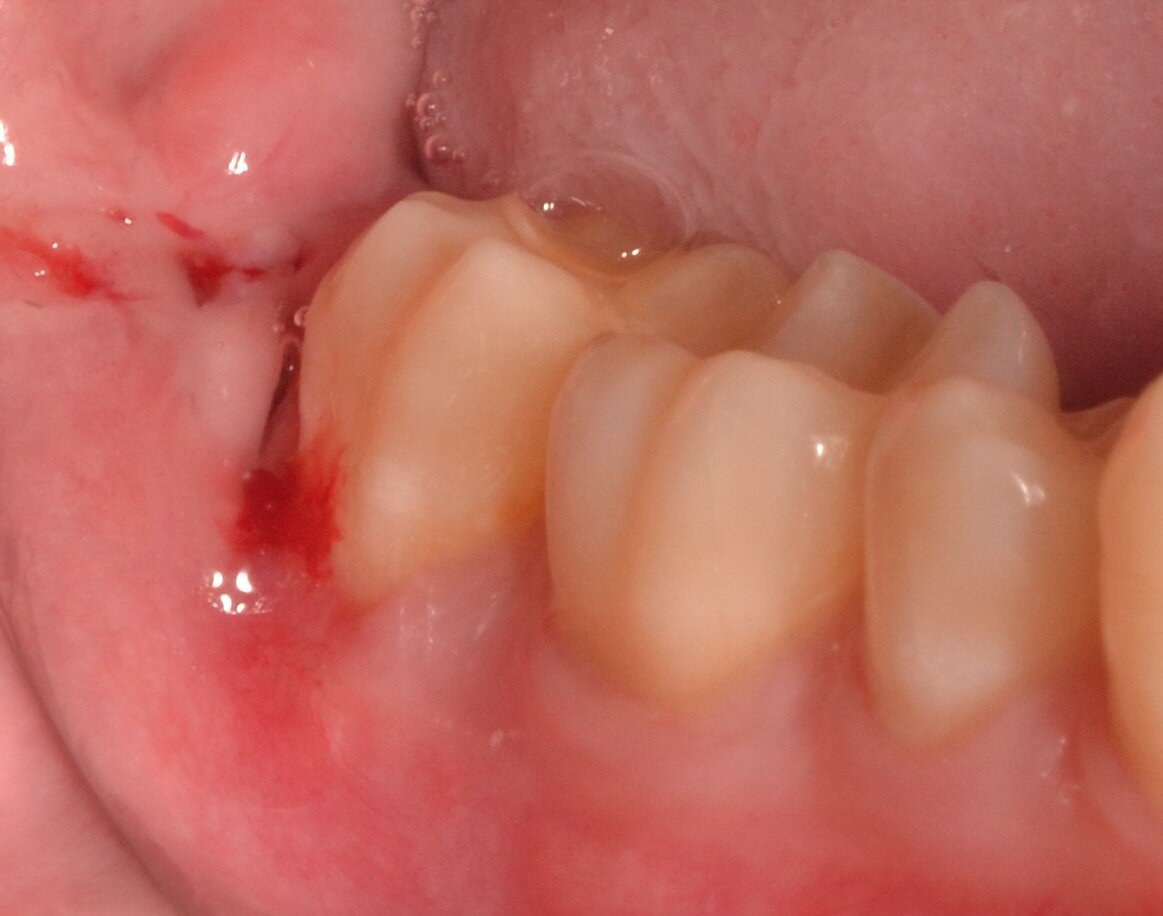
In the oral cavity, tooth 4.8 has erupted with mesial cusps, has a vertical position and a vestibular inclination. The surrounding mucosa shows no signs of inflammation, and palpation is painless (see Fig. 1A). On CBCT the tooth shows signs of a vertical position, vestibular inclination, signs of tooth root penetration into the mandibular canal and interruption of its cortical plate, which corresponds to a high risk of trauma to the mandibular nerve in case of complete extraction of tooth 4.8 (Fig. 2).
A decision was made to perform a coronectomy of tooth 4.8, which was carried out in accordance with the above-described technique (see Fig. 1).
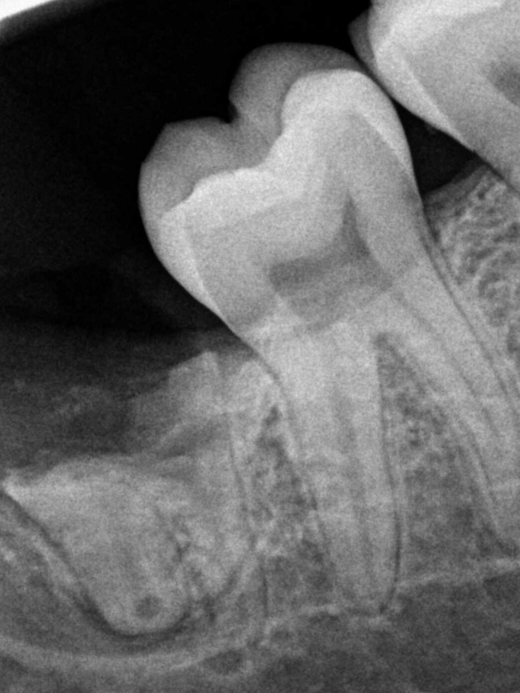
Immediately after the operation, the periapical radiograph showed signs of the removed crown of tooth 4.8, with the position of the tooth roots unchanged. The coronal border of the remaining roots is below/at the level of the surrounding crestal bone (Fig. 3).
The postoperative period was uneventful. An examination was performed 7 days later for removing the sutures. The patient complained of mild pain in the postoperative area. The postoperative area was in the epithelialization stage, palpation was painless, the sutures were loosened, and the discharge from the wound was serous (Fig. 4).
At a follow-up examination 3 months after surgery the surrounding mucosa showed no signs of inflammation, palpation was painless, and there were no signs of eruption of the roots. According to CBCT data, signs of “eruption” of the roots of tooth 4.8 and a change in the relative position of the apices of the tooth roots and the mandibular canal were found. A minimal distance from the roots of tooth 4.8 to the CEC line of tooth 4.7 was noted (Fig. 5). Despite a small area of enamel found in the distal part of the tooth root, this did not affect the overall clinical and radiographic picture.
One and a half years after coronectomy, the clinical picture is similar to that observed three months after the procedure. The CBCT shows signs of continued “eruption” of the roots of tooth 4.8. No signs of pathological changes in the bone tissue were found (Fig. 6).
 A
A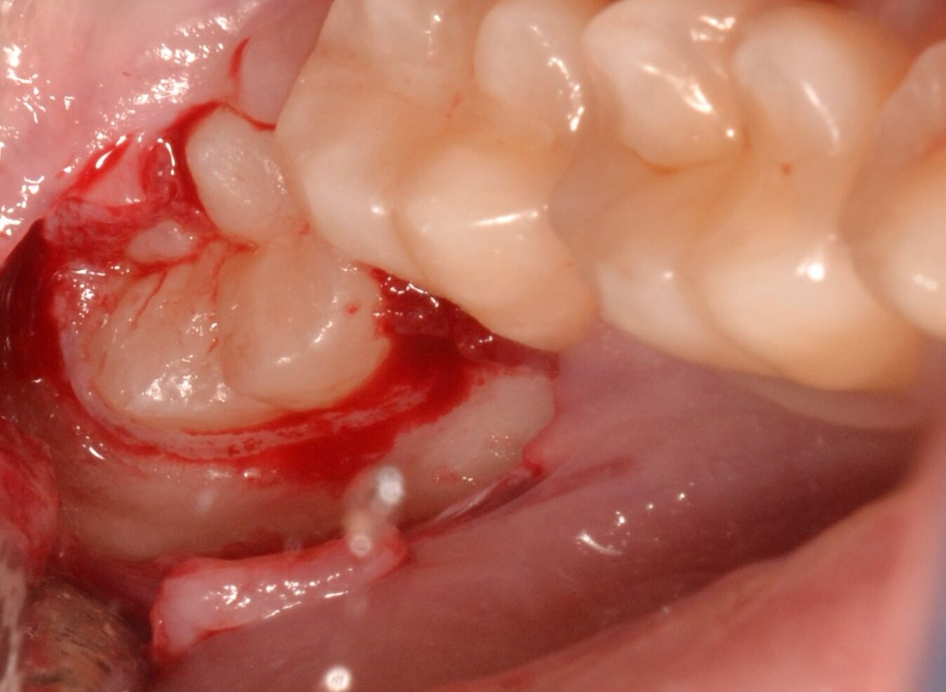 B
B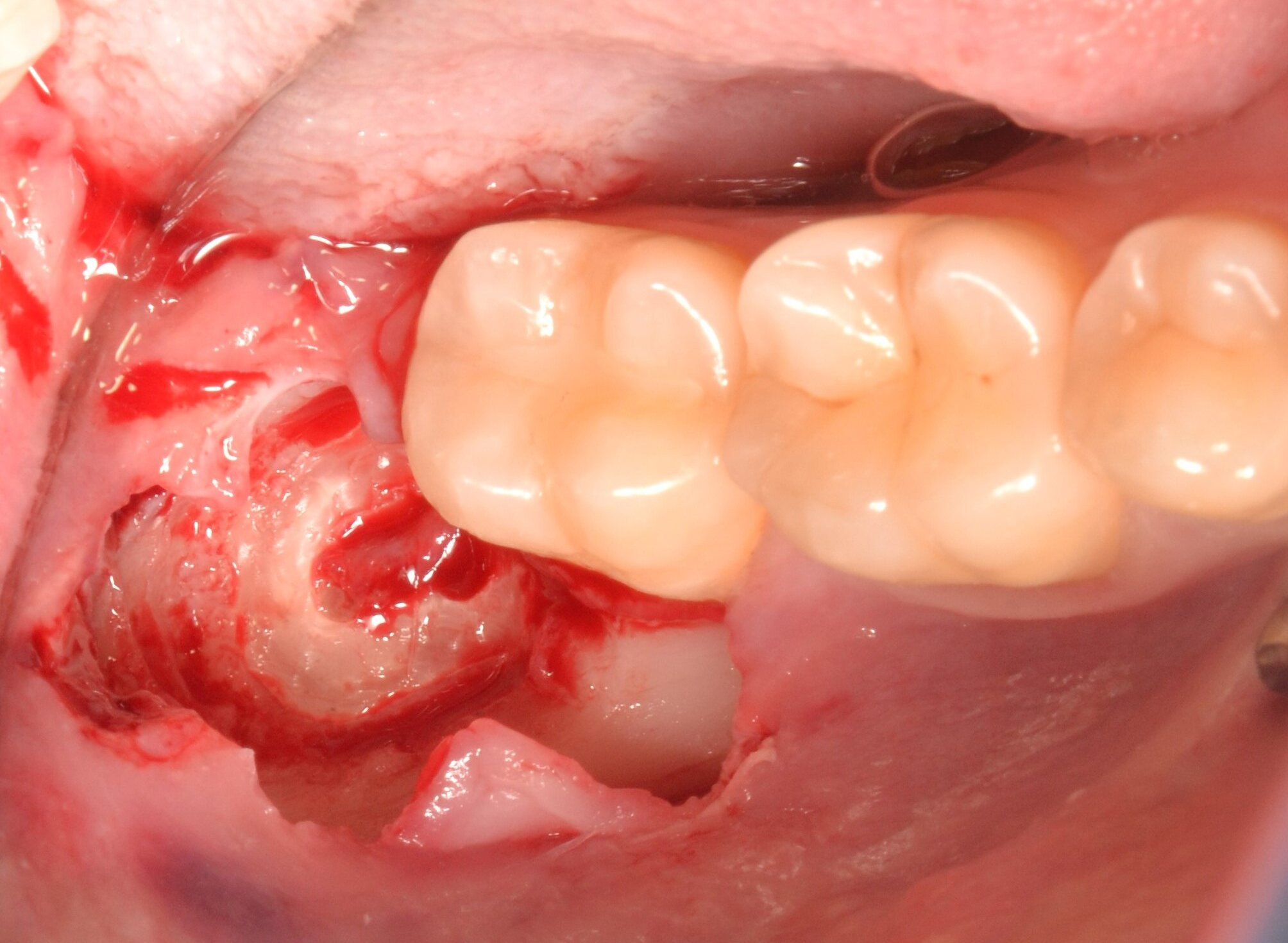 C
C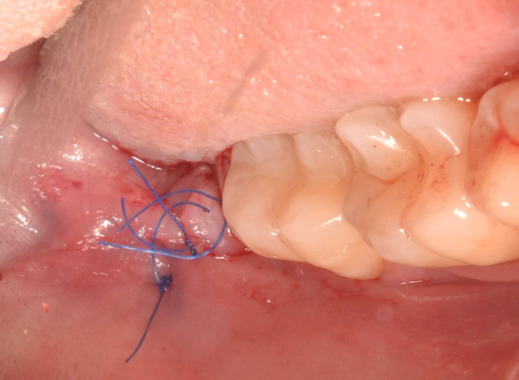 D
D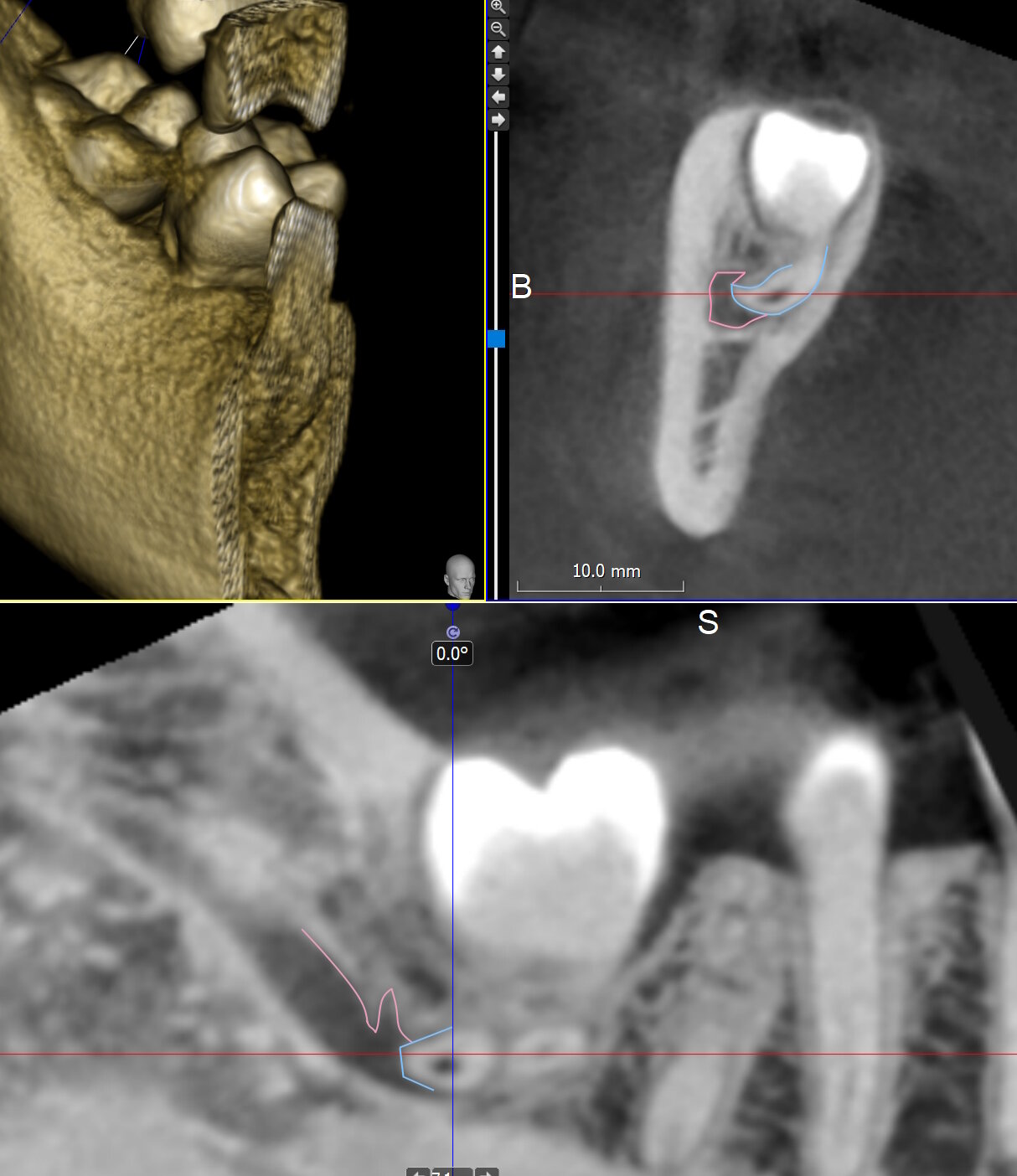 A
B
C
A
B
C
 A
A B
B C
C A
A B
B C
CClinical case 2
Patient D., 27 years old, was referred for extraction of tooth 3.8 prior to orthodontic treatment.
Tooth 3.8 is not visible in the oral cavity, the surrounding mucosa shows no signs of inflammation, palpation is painless. On CBCT, tooth 3.8 shows signs of vertical position, signs of the tooth roots enveloping the mandibular canal, which corresponds to a high risk of trauma to the mandibular nerve in case of complete tooth extraction. A decision was made to perform a coronectomy of tooth 3.8.
Immediately after the coronectomy, signs of the removed crown of tooth 3.8 are determined on the CBCT, the position of the tooth roots is unchanged. The coronal border of the remaining roots is below the surrounding crestal bone. An area of enamel was found in the distal part of the tooth, which was immediately removed. The minimum distance from the coronectomy border to the CEJ of tooth 3.7 was measured (Fig. 7).
The postoperative period was uneventful. After 7 days, the patient complained of mild pain in the postoperative area when eating hot or cold food. On examination: the postoperative area was in the epithelialization stage, palpation was painless, and the sutures were loosened. The sutures were removed.
At a follow-up examination 10 months later, the gingiva of the retromolar area showed no visible pathological changes, palpation was painless, and there were no signs of eruption of the roots of tooth 3.8 through the gingiva (Fig. 8). The patient is undergoing orthodontic treatment. A follow-up CBCT scan showed signs of “eruption” of the roots of tooth 3.8, signs of native bone tissue formation coronal to the roots of tooth 3.8, and signs of pathological changes.
At the follow-up examination 2 years after coronectomy, the postoperative area showed no visible pathological changes, palpation was painless, and there were no signs of tooth root eruption through the mucous membrane. The follow-up CBCT showed signs of continued “eruption” of the roots of tooth 3.8, with no signs of pathological changes in the bone tissue (Fig. 9).
 A
A B
B C
C A
A B
B C
C A
A B
B C
CClinical case 3
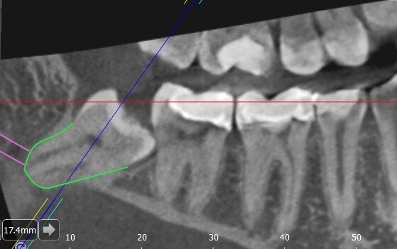
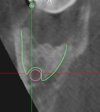
Patient D., 26 years old, referred for extraction of tooth 4.8 prior to orthodontic treatment. Tooth 4.8 is not visible in the oral cavity, the surrounding mucosa is painless with no signs of inflammation. On CBCT, tooth 4.8 showed signs of mesial inclination and signs of “envelopment” of the mandibular canal by the tooth roots, which corresponds to a high risk of trauma to the mandibular nerve in case of complete extraction of tooth 4.8 (Fig. 10). A decision was made to perform a coronectomy.
The surgical protocol was performed in accordance with the above-described technique (Fig. 11). A postoperative CBCT was performed to check the quality of the coronectomy, and the minimum distance from the roots of tooth 4.8 to the CEC of tooth 4.7 was also measured (Fig. 12).
The postoperative period proceeded without complications. An examination was performed seven days later, and the sutures were removed. The patient had no complaints. The postoperative area was in the epithelialization stage, palpation was painless, and the sutures were loosened. The sutures were removed.
At a follow-up examination 4 months after coronectomy, the gingiva of the retromolar area showed no visible pathological changes, palpation was painless, and there were no signs of tooth root eruption through the gingiva. The patient is undergoing orthodontic treatment. At the follow-up CBCT, no signs of “eruption” of the roots of tooth 4.8 or pathological changes in the bone tissue were found, and the minimum distance from the roots of tooth 4.8 to the center of the crown of tooth 4.7 was measured (Fig. 13).
At the follow-up examination 1 year after coronectomy, the mucosa of the retromolar region showed no visible pathological changes, palpation was painless, and no signs of eruption of the roots of tooth 4.8 through the gingiva were found. On follow-up CBCT, there were signs of continued “eruption” of the roots of tooth 4.8 with no pathological changes in the bone tissue (Fig. 14).
 A
A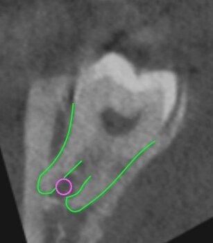 B
B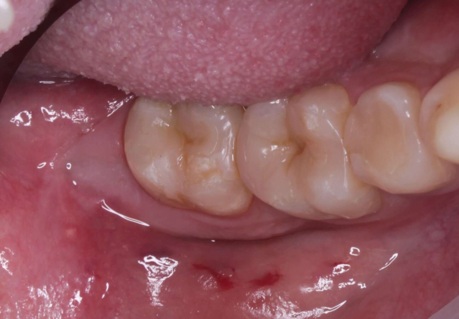 A
A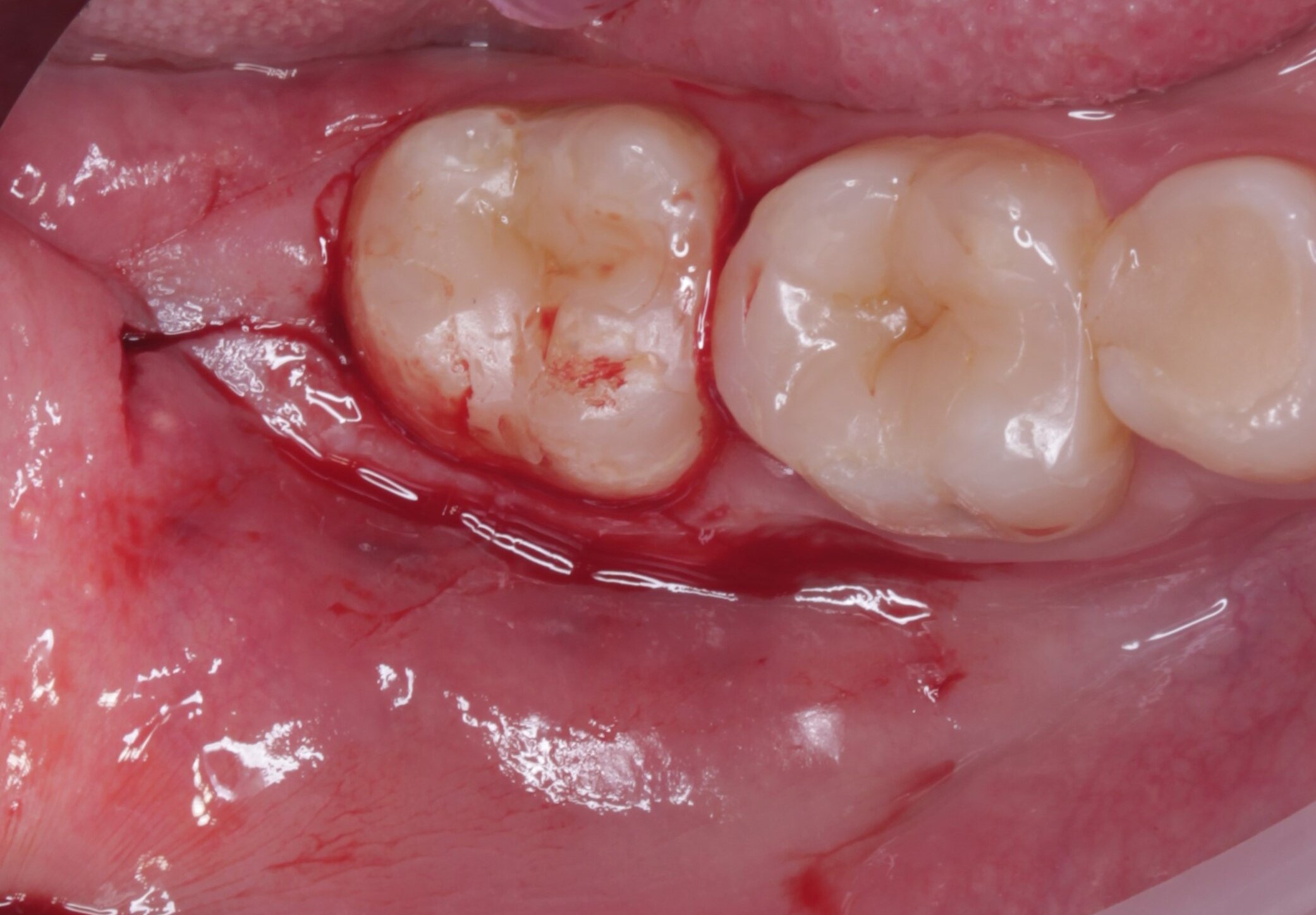 B
B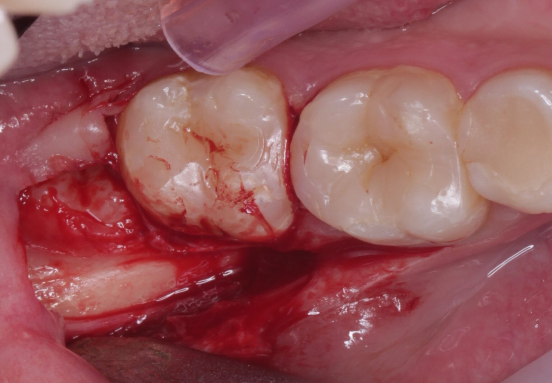 C
C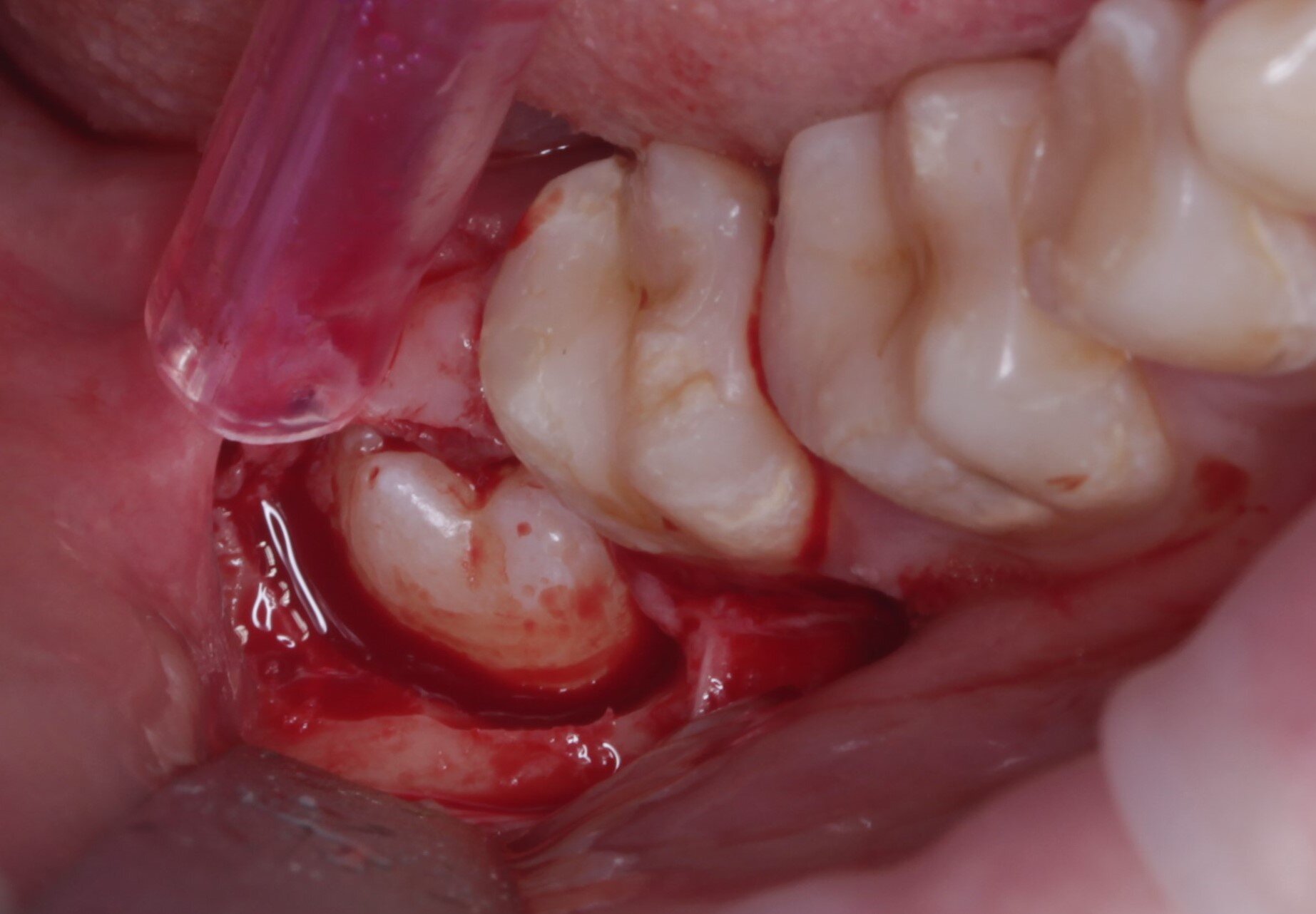 D
D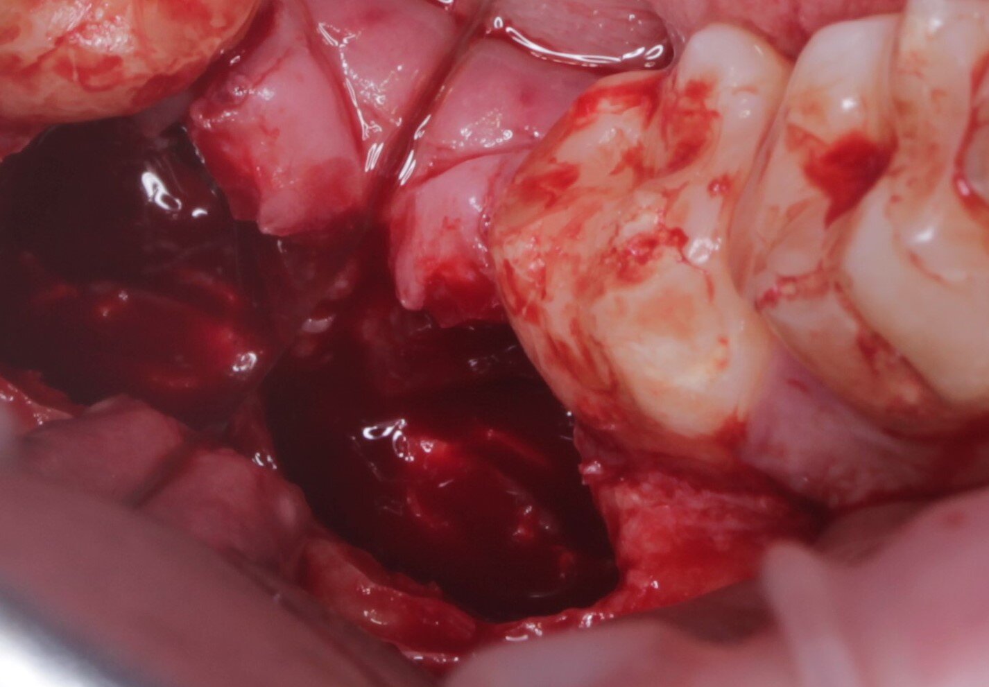 E
E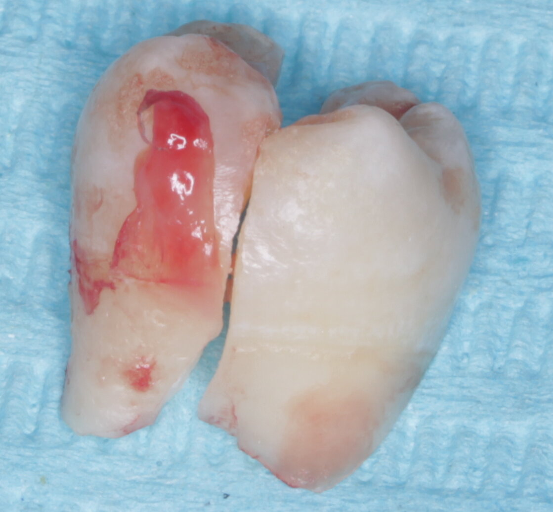 G
G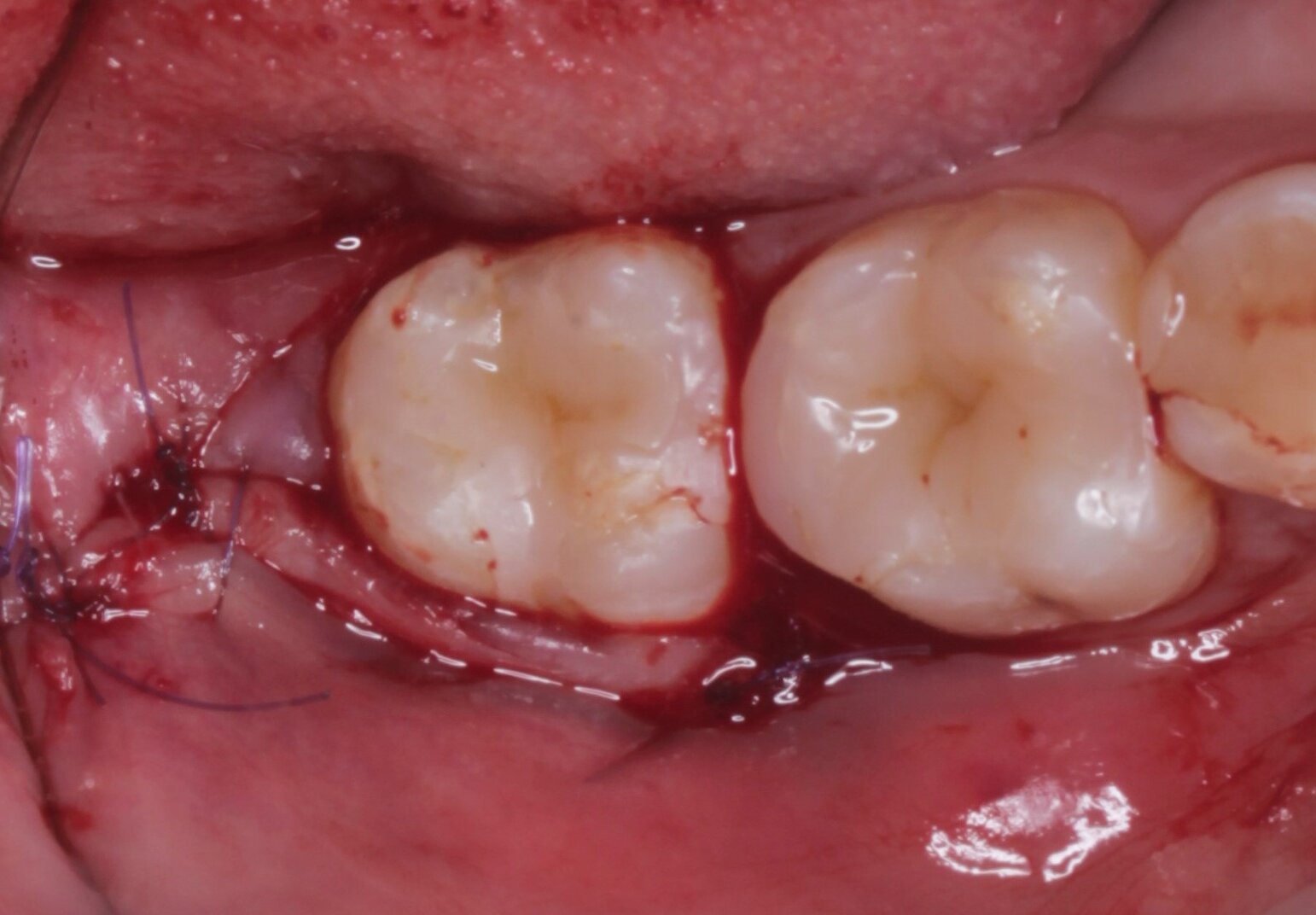 H
H
 A
A B
B C
C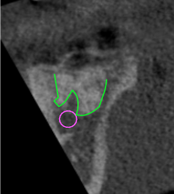 A
A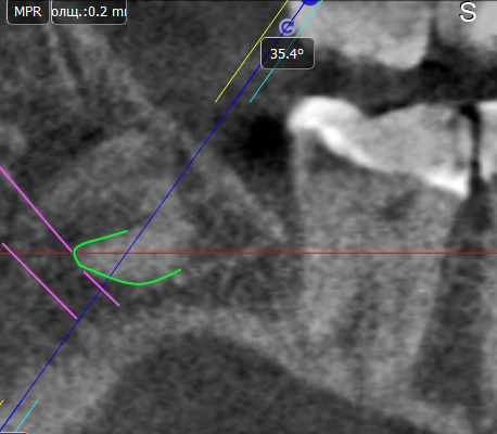 B
B C
CDiscussion
The success of coronectomy is not accidental, it is based on biological principles previously proven by authors of scientific papers. For example, histological and biochemical studies of the composition and structure of dentin, cementum, and bone tissue revealed similarities [20, 21], which allow these structures to fuse with each other, for example, in cases of tooth ankylosis, and also allow dentin to be replaced by bone tissue, for example, in cases of replacement resorption without the development of inflammatory reactions [22, 23]. This fact also has great practical significance: dentin blocks or dentin autografts are used as bone grafting material to replace bone tissue defects with good clinical and morphological results [24, 25]. Therefore, in the vast majority of situations, the presence of intact roots in bone tissue does not cause a reactive response from the body.
Enamel, a structure different from dentin, requires complete extraction from the surgical area whenever possible, as it cannot be replaced by bone tissue and periodontal fibers do not attach to it as they do to root cementum. For example, when forming dentin autoblocks or preparing dentin autografts, the crown part of the tooth with enamel is removed [26–28].
The roots of the tooth remaining in the socket after coronectomy do not require endodontic treatment. It has been shown that endodontic treatment reduces the success of coronectomy [29]. Presumably, aseptic inflammation occurs in the superficial areas of the root pulp at the border of the coronectomy zone due to thermal burns caused by rotating diamond instruments, followed by revascularization of the pulp due to the formation of a blood clot in the socket as well as preservation of the neurovascular bundle in the apical part of the tooth. This phenomenon has the same biological principles and is similar to the clinical situation when apicoectomy of the donor tooth is performed during autotransplantation of a tooth with a formed root apex to preserve the vitality of the tooth in the recipient area [30]. Histological examination of tooth roots in unsuccessful coronectomy operations showed signs of pulp tissue vitality in such roots, as well as the absence of signs of perirradicular inflammation [31].
At the same time, it cannot be said that coronectomy surgery is free from possible complications. The risks of soft tissue trauma increase in cases of deep tooth retention, as well as when the tooth is tilted lingually, since adequate visualization of the working field is necessary for complete extraction of the tooth crown enamel to the level of the CEJ, which is difficult to achieve in such a clinical situation and may require the detachment of a full-thickness flap from the lingual side, thereby increasing the risk of trauma to the lingual nerve.
Dalle Carbonare et al. (2017) presented a systematic review describing cases of identified signs of trauma to the inferior alveolar nerve and lingual nerve in both successful and unsuccessful coronectomies. Of the 2,087 coronectomy operations, 1,935 were successful and 152 were unsuccessful. In successful coronectomies, signs of trauma to the IAN were detected in 10 cases out of 1,935 (0.5%), of which 1 case (0.05%) had permanent paresthesia. In cases of unsuccessful coronectomy, signs of trauma to the lingual nerve were found in 4 out of 152 cases (3%), of which in 2 cases (1.3%) the paresthesia was permanent. Damage to the lingual nerve was described in 1 case (0.05%) of successful coronectomy and in none of the unsuccessful cases. The reasons for unsuccessful coronectomy included: pulpitis 4/152 (3%), uncut enamel areas 10/152 (7%), wound edge divergence 14/152 (9%), infection 12/152 (8%), root migration and eruption 50/152 (33%), root mobility 55/152 (36%), unknown causes 4/152 (3%), and others 3/152 (2%; chronic periodontitis, gingival hyperplasia distal to the second molar, or orthognathic surgery) [32].
In cases of horizontal retention and when the IAN is close to the crown of the third molar, and in situations where maximum precision in controlling the depth of immersion of the rotating instrument is important, the authors recommend using marked surgical burs [33], which allow avoiding trauma to the structures of the mandibular canal and reducing operating time, ensuring convenience and confidence in work. If this is not possible, stepwise preparation with depth control using a periodontal graduated probe is strongly recommended.
In addition to coronectomy, there are other techniques for removing lower third molars whose roots are in direct contact with the IAN. One such technique is orthodontic extrusion of lower third molars followed by their complete extraction [34]. This technique allows avoiding trauma to the mandibular nerve during deep tooth retention, since after extrusion, the distance between the mandibular nerve and the tooth root increases, the tooth itself becomes mobile, and, accordingly, the operation of complete tooth extraction becomes easier manually and safer in terms of the risks of mandibular nerve trauma. This technique also avoids bone loss on the distal side of the second lower molar of the lower jaw due to the extrusion of the wisdom tooth. However, it should be noted that this method is not without its drawbacks, among which the following can be highlighted: the need to fix the orthodontic structure, the accompanying discomfort from trauma to the mucous membrane due to the limited space in the retromolar area. There is also a high probability of repositioning of mini-screws and orthodontic appliances on the tooth crown due to mobility and adhesion problems, increased treatment time, and multiple visits to the orthodontist for treatment monitoring and replacement of elastic ties. Another disadvantage is the fact that fixing the orthodontic structure requires detaching the flap, exposing the tooth crown, keeping the surgical field dry, and, in general, creating a space in the oral cavity where the extrusion will be performed. At the same time, this technique may be an option in cases where it is difficult to perform a coronectomy.
In addition to orthodontic extrusion, foreign literature also describes the method of pericoronal osteotomy [35], which involves removing bone tissue around the crown of the impacted tooth, as well as its slight luxation, in order to stimulate the process of its eruption. In this case, repeated surgical intervention is also required after 6–8 weeks for complete extraction of the tooth after its coronal displacement. It should be noted that this method is only suitable if there is an optimal eruption vector (e.g., a vertically positioned tooth or a tooth with a slight medial inclination). Otherwise, the space for eruption will be limited by the second molar or completely impossible due to the horizontal position of the third molar, which greatly limits the application of this technique.
In addition to cases of third molar coronectomy, options for coronectomy of other groups of teeth are also described with the aim of preserving the volume of the alveolar ridge before prosthetics [36]. The section of the tooth root left in the bone minimizes bone loss in this area, an example of which is the “vestibular shield” technique in dental implantation [37].
The question of whether to perform a coronectomy on asymptomatic lower third molars with a vertical position remains debatable in cases where patients are referred by an orthodontist for complete tooth extraction to allow for distal movement of the teeth. In fact, performing a coronectomy on such teeth does not free up space in the jawbone distal to the second molar, and this surgical approach may affect the final outcome of orthodontic treatment. In such cases, we believe that complete extraction of the third molar can be performed in two stages: the first stage is coronectomy, followed by “eruption” of the tooth roots approximately 6 months later; and upon detection of signs of improved alignment between the tooth roots and the alveolar bone following a repeat CBCT scan, complete extraction of the tooth roots is performed.
Clinical cases have demonstrated the possibility of coronal migration of tooth roots following coronectomy, or so-called “eruption.” On the one hand, this can be a negative factor, as when tooth roots erupt through the alveolar mucosa, a repeat surgery for complete root removal may be required. On the other hand, anatomically, such roots will be located at a distance from the inferior alveolar nerve (IAN), and their removal will be significantly simpler compared to the initial situation and will not be associated with the risk of paresthesia.
However, despite the positive results in the above clinical cases, this study has limitations related to the small number of detailed clinical cases and the observation period (less than 5 years). More studies with a longer observation period are needed on coronectomy, as this technique is still experimental in the domestic dental community. The phenomenon of root migration requires detailed study, namely the influence of the distance from the coronal border of the remaining roots to the edge of the alveolar process and the morphology of the roots on the magnitude and speed of migration, the formation of bone tissue over the roots of the teeth after coronectomy, and the frequency and causes of eruption of the remaining roots through the mucous membrane of the retromolar region. The issue of developing a classification of the degree of risk of complications associated with damage to the IAN during the extraction of the lower third molar remains unresolved in order to determine clear indications for coronectomy.
Conclusion
Based on the clinical and radiographic picture of the long-term results of the presented clinical cases, the authors conclude that coronectomy is a reliable alternative to the classic complete extraction of vital asymptomatic lower third molars, which is associated with a high risk of trauma to the IAN. The authors recommend coronectomy as a full-fledged option for surgical treatment in cases with a high risk of trauma, or as the first stage followed by complete extraction of the tooth after it has “erupted.”
References
- Kiencało A., Jamka-Kasprzyk M., Panaś M., Wyszyńska-Pawelec G. Analysis of complications after the removal of 339 third molars. Dent Med Probl. 2021; 58 (1): 75—80. PMID: 33789003
- Ghaeminia H., Nienhuijs M.E., Toedtling V., Perry J., Tummers M., Hoppenreijs T.J., Van der Sanden W.J., Mettes T.G. Surgical removal versus retention for the management of asymptomatic disease-free impacted wisdom teeth. Cochrane Database Syst Rev. 2020; 5 (5): CD003879. PMID: 32368796
- Chaddoud M., Anokhina A.V. Modern views on the problem of preserving the third permanent molars (review of publications). Acta Medica Eurasica. 2024; 2: 86—96 (In Russian). DOI: 10.47026/2413-4864-2024-2-86-96
- Baibakov S.E., Bakhareva N.S., Dorogan V.V., Dorogan V.V. Structure characteristics of mandibular canal based on cone beam computed tomography. International Research Journal. 2021; 6—2 (108): 98—102 (In Russian). DOI: 10.23670/IRJ.2021.108.6.052
- Achararit P., Manaspon C., Jongwannasiri C., Kulthanaamondhita P., Itthichaisri C., Chantarangsu S., Osathanon T., Phattarataratip E., Sappayatosok K. Impacted lower third molar classification and difficulty index assessment: comparisons among dental students, general practitioners and deep learning model assistance. BMC Oral Health. 2025; 25 (1): 152. PMID: 39875882
- Cheung L.K., Leung Y.Y., Chow L.K., Wong M.C., Chan E.K., Fok Y.H. Incidence of neurosensory deficits and recovery after lower third molar surgery: a prospective clinical study of 4338 cases. Int J Oral Maxillofac Surg. 2010; 39 (4): 320—6. PMID: 20061121
- Haug R.H., Perrott D.H., Gonzalez M.L., Talwar R.M. The American Association of Oral and Maxillofacial Surgeons age-related third molar study. J Oral Maxillofac Surg. 2005; 63 (8): 1106—14. PMID: 16094577
- Kuzin A.V., Sogacheva V.V., Gurin A.N., Syomkin V.A., Sogachev G.V., Remizov G.V. Surgical treatment tactics in patients with third molars located close to mandibular canal. Stomatology. 2022; 6: 73—78 (In Russian). DOI: 10.17116/stomat202210106173
- Lezhenina S.V., Kuzmina A.A. Damage to lower alveolar vascular bundle. Neuropraxia, axonomotis, neurotomesis. 7 p. (In Russian). eLIBRARY ID: 67334047
- Coulthard P., Kushnerev E., Yates J.M., Walsh T., Patel N., Bailey E., Renton T.F. Interventions for iatrogenic inferior alveolar and lingual nerve injury. Cochrane Database Syst Rev. 2014; 2014 (4): CD005293. PMID: 24740534
- Diker N., Caglayan B., Helvacioglu F., Kilic E. The effect of systemic rifampicin treatment on inferior alveolar nerve regeneration in rats following crush injury. Eur J Oral Sci. 2020; 128 (3): 183—189. PMID: 32236990
- AlAli A.M., AlAnzi T.H. Inferior alveolar nerve damage secondary to orthodontic treatment: A systematic scoping review. Int J Risk Saf Med. 2021; 32 (3): 175—191. PMID: 33579879
- Ma Y., Yang M., Chen X., Qu W., Qu X., He P. The effectiveness of photobiomodulation therapy on inferior alveolar nerve injury: A systematic review and meta-analysis. PLoS One. 2023; 18 (8): e0287833. PMID: 37561792
- Nayak V., et al. Association of radiographic signs in determining the proximity of mandibular third molar roots to the mandibular canal and postoperative occurrence of neurosensory disorders: A cohort study. Cureus. 2023; 15 (12): e51085. PMID: 38274929
- Goryacheva E.V., Zyul’kina L.A., Koretskaya E.A., Chesnykh E.A., Kofova T.E. The morphological features of the mandibular canal position in relation to the third molars’ roots (a review of literature). University Proceedings. Volga region. Medical sciences. 2021; 3 (59): 77—89 (In Russian). DOI: 10.21685/2072-3032-2021-3-8
- Grigoryants A.G., Khatypova A.E., Makalish T.P., Kutya S.A. Applied anatomy of mandibular canal. Humans and their health. 2023; 3: 69—75 (In Russian). DOI: 10.21626/vestnik/2023-3/08
- Kuzin A.V., Vedyaeva A.P., Sogachyova V.V., Gurin A.N., Remizov G.V. Microanatomy features in third molars roots adjacent to mandibular canal. Stomatology. 2023; 6: 16—21 (In Russian). DOI: 10.17116/stomat202310206116
- Badalyan V.A., Drobot G.V., Brutian V.A., Kopilov M.V. Method for removal of impacted third molars in close proximity with the lower alveolar nerve. Clinical Dentistry (Russia). 2019; 2 (90): 58—61 (In Russian). DOI: 10.37988/1811-153X_2019_2_58
- Williams M., Tollervey D. Lower third molar surgery — consent and coronectomy. Br Dent J. 2016; 220 (6): 287—8. PMID: 27012340
- Feng Y., Zhao R., Li J., Yuan Z., Xu X., Gong J. Efficacy of autogenous particulated dentin graft for alveolar ridge preservation: A systematic review and meta-analysis of randomized controlled trials. Medicine (Baltimore). 2023; 102 (48): e36391. PMID: 38050282
- Yüceer-Çetiner E., Özkan N., Önger M.E. Effect of autogenous dentin graft on new bone formation. J Craniofac Surg. 2021; 32 (4): 1354—1360. PMID: 33405453
- Hanisch M., Hanisch L., Kleinheinz J., Jung S. Primary failure of eruption (PFE): a systematic review. Head Face Med. 2018; 14 (1): 5. PMID: 29544499
- Tsukiboshi M., Yamauchi N., Tsukiboshi Y. Long-term outcomes of autotransplantation of teeth: A case series. J Endod. 2019; 45 (12S): S72-S83. PMID: 31623912
- Elraee L., Abdel Gaber H.K., Elsayed H.H., Adel-Khattab D. Autogenous dentin block versus bone block for horizontal alveolar ridge augmentation and staged implant placement: A randomized controlled clinical trial including histologic assessment. Clin Oral Implants Res. 2022; 33 (7): 723—734. PMID: 35509125
- Sidorenko V.O., Panin A.M., Ciciashvili A.M., Gabidullina V.R. Alveolar bone plasty using removed teeth autodentine possibility analysis. Russian Stomatology. 2019; 3: 14—18 (In Russian). DOI: 10.17116/rosstomat20191203114
- Speranskaya E.M., Golubtsova N.N., Mukhamedzhanova L.R., Moskvichev E.V. E-cadherin and the gingival epithelial barrier. Medical News of North Caucasus. 2022; 4: 461—466 (In Russian). DOI: 10.14300/mnnc.2022.17111
- Zhang S., Li X., Qi Y., Ma X., Qiao S., Cai H., Zhao B.C., Jiang H.B., Lee E.S. Comparison of autogenous tooth materials and other bone grafts. Tissue Eng Regen Med. 2021; 18 (3): 327—341. PMID: 33929713
- Grawish M.E., Grawish L.M., Grawish H.M., Grawish M.M., Holiel A.A., Sultan N., El-Negoly S.A. Demineralized dentin matrix for dental and alveolar bone tissues regeneration: An innovative scope review. Tissue Eng Regen Med. 2022; 19 (4): 687—701. PMID: 35429315
- Sencimen M., Ortakoglu K., Aydin C., Aydintug Y.S., Ozyigit A., Ozen T., Gunaydin Y. Is endodontic treatment necessary during coronectomy procedure? —J Oral Maxillofac Surg. 2010; 68 (10): 2385—90. PMID: 20663600
- Rugani P., Kirnbauer B., Mischak I., Ebeleseder K., Jakse N. Extraoral root-end resection may promote pulpal revascularization in autotransplanted mature teeth—A retrospective study. J Clin Med. 2022; 11 (23): 7199. PMID: 36498773
- Patel V., Sproat C., Kwok J., Beneng K., Thavaraj S., McGurk M. Histological evaluation of mandibular third molar roots retrieved after coronectomy. Br J Oral Maxillofac Surg. 2014; 52 (5): 415—9. PMID: 24684971
- Dalle Carbonare M., Zavattini A., Duncan M., Williams M., Moody A. Injury to the inferior alveolar and lingual nerves in successful and failed coronectomies: systematic review. Br J Oral Maxillofac Surg. 2017; 55 (9): 892—898. PMID: 29061470
- Smirnov A.D., Yusaev T.A. Case report of impacted mandibular third molar extraction using marked surgical bur. Stomatology. 2022; 5: 73—76 (In Russian). DOI: 10.17116/stomat202210105173
- Alessandri Bonetti G., Bendandi M., Laino L., Checchi V., Checchi L. Orthodontic extraction: riskless extraction of impacted lower third molars close to the mandibular canal. J Oral Maxillofac Surg. 2007; 65 (12): 2580—6. PMID: 18022488
- Tolstunov L., Javid B., Keyes L., Nattestad A. Pericoronal ostectomy: an alternative surgical technique for management of mandibular third molars in close proximity to the inferior alveolar nerve. J Oral Maxillofac Surg. 2011; 69 (7): 1858—66. PMID: 21549479
- Mahesh L., Bhasin M.T. Root submergence technique, a case series with follow-up up to seven years. Acta Scientific Dental Sciences. 2022: 6 (9): 55—60. DOI: 10.31080/ASDS.2022.06.1454
- Ogawa T., Sitalaksmi R.M., Miyashita M., Maekawa K., Ryu M., Kimura-Ono A., Suganuma T., Kikutani T., Fujisawa M., Tamaki K., Kuboki T. Effectiveness of the socket shield technique in dental implant: A systematic review. J Prosthodont Res. 2022; 66 (1): 12—18. PMID: 33692284