DOI:
10.37988/1811-153X_2021_4_92Combined use of 2D- and 3D-simulation results for identical smile prototype production (clinical case)
Downloads
Abstract
The article presents a clinical case of using the result of converting a smile 2D-design into a 3D-treatment scene and making an identical prototype of future restorations.Materials and methods.
To achieve the goal, the Avantis 3D program was used, into which the results of the smile 2D-design were imported and a 3D-treatment scene was subsequently created. Results and discussion. According to the result of making virtual and physical prototypes of a future smile, it was possible to recreate identical shapes and size of teeth agreed with the patient at the stage of 2D design.
Key words:
2D-treatment planning, 3D-treatment planning, Avantis 3D, smile design, 3D-printer, digital dentistryFor Citation
Modern aesthetic dentistry includes digital diagnostic tools, planning and visualization of future dentures using computer modeling and manufacturing methods. Important stages preceding orthopedic rehabilitation in an aesthetically significant area are 2D design and 3D modeling of the patient's future smile to harmonize the main parameters of shape, color, and position of teeth in the dentition [1]. This approach minimizes possible conflict situations associated with the patient's dissatisfaction with the final result of prosthetics, and allows all specialists involved in the process to predict the outcome of treatment [2].
Today there are many ways of how to make a smile using virtual planning. These can be basically divided into two groups — 2D design and 3D design [3].
The advantages of 2D design include simplicity and an ability to use the software not only by the dentist, but also by the patient, which makes it possible to carry out the preliminary 2D planning stage even before visiting the dental clinic. However, an important disadvantage of such modeling is the presence of the initial data in only one projection, which complicates the further reproduction of restorations by a dental technician [4].
3D smile design allows not only obtaining full-fledged 3D data about a future smile, but also makes it possible to accurately reproduce the resulting virtual structures by milling or prototyping.
The complexity of using 3D modeling programs is associated with the high time spent on design, the inaccessibility of software for patients, as well as the difficult mastering of various 3D programs by dentists and dental technicians [5].
The most popular tool in 2D smile design has been DSD (Digital Smile Design) technology for many years. The design of a future smile in this program can be seen using processed digital photographs. Another important point is the innovative method of aesthetic and clinical planning in aesthetic and prosthetic dentistry, which is of great importance for the analysis and design in the dental laboratory. This technique can be also used for diagnostics and planning in plastic and maxillofacial surgery. First of all, the protocol provides for the acquisition of images of the patient through digital photographs and digital video filming. The video is very important — you can see the dynamic phases of a smile associated with physiological characteristics (facial expressions, phonetics, the ratio of the dentition and lips). Recording this important information on the patient's digital aesthetic card complements the anamnesis as it is an integral part of the objective intra- and extraoral examination as well as the aesthetic analysis.
The main drawback of programs for 2D smile design is an inaccuracy of the reproduced physical models of teeth, considering the human factor which can be resulted in a patient’s dissatisfaction in dental rehabilitation. In comparison to 2D design, 3D design supposed to have no drawback at all [6].
The possibility of converting a patient-approved 2D smile design into a 3D format is an urgent task of modern digital dentistry, and the combined use of two technologies determined the purpose of the study [7].
The aim of the study is to develop a technique that ensures the accuracy of reproducing a virtual 2D prototype of a smile when restoring a patient's teeth through the combined use of 3D modeling methods.
Materials and methods
To achieve this goal, we proposed a method for the combined use of 2D and 3D modeling programs. With an intraoral scanner, optical impressions of the dentition of the upper and lower jaw were obtained from the patient and the bite was recorded.
- Take a full-face portrait photograph of the patient with a wide smile.
- Based on the photograph received on the computer in the SmileCloud online service, the doctor conducts a 2D model of the smile, selecting the shape and position of the teeth, observing the rules of aesthetic symmetry, so that the image of the gingival papillae of the teeth located in the smile zone is preserved.
- The resulting 2D design of the smile was agreed with the patient.
- The obtained scans of the jaws and the photograph with the models of the teeth in the smile zone were loaded into the program for modeling dentures Avantis 3D (Avantis 3D, Russia).
- By the points symmetrically set on the tops of the gingival papillae of the teeth located in the smile zone, volumetric images of the teeth were obtained, which must be obtained by the method of intraoral 3D scanning, comparing with the patient's photograph.
- On top of the virtual volumetric image of the patient's teeth, the shape of future dentures was simulated using the electronic library of teeth, the most suitable in shape to the 2D design agreed with the patient.
- After the completion of the modeling, the models of jaws with artificial teeth were made using the 3D-printing method.
The difficulty of comparing 2D and 3D images in the Avantis 3D program for modeling dentures is that symmetrical points must be set on the most geometrically selected objects of the teeth, for example, along the medial angle of the clinical crown of the incisor. And this is impossible, given that the image of the teeth in the plane photo is covered by virtual models.
In the proposed method, this problem is solved by comparing the images by symmetric points installed on the tops of the gingival papillae shown in the planar photograph and the volumetric image of the jaws obtained by the method of intraoral scanning.
When translating 2D design results into 3D modeling, the transparency adjustment function is used to control the identity of the alignment of the two images.
Results and discussion
As a result of the clinical effectiveness of the proposed method for virtual modeling of prototypes of future dentures, we present a clinical example. Patient J., 25 years old, came to the clinic with complaints of an aesthetic defect in the anterior teeth of the upper jaw. After diagnostic measures, it was decided to manufacture ceramic veneers with support on teeth 1.3, 1.2, 1.1, 2.1, 2.2 and 2.3.
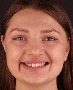
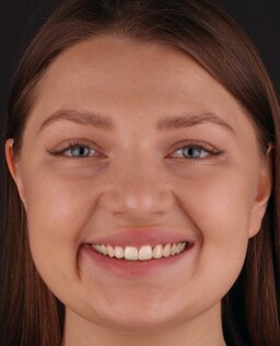
The patient underwent intraoral scanning, optical impressions of the upper and lower dentition were obtained with registration of the bite. We also obtained a full-face portrait photograph with a wide smile, which was used for 2D mock-up of the smile in the SmileCloud program (Fig. 1).
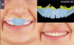
The obtained images were loaded into the Avantis 3D software, in which the three-dimensional image of the maxillary teeth was compared with the photographs of the patient with a wide smile using the points symmetrically set on the apices of the gingival papillae of teeth 1.3, 1.2, 1.1, 2.1, 2.2 and 2.3. Above the virtual volumetric image of teeth 1.3, 1.2, 1.1, 2.1, 2.2 and 2.3, volumetric modeling of the prototype of future veneers was carried out (Fig. 2).
For this purpose, we used the electronic library of teeth available in the program and the most suitable for the planar model agreed with the patient. After that, the shapes of the vestibular surfaces of the teeth 1.3, 1.2, 1.1, 2.1, 2.2 and 2.3 of the modeled volumetric prototype of the denture designs were adapted to the similar surfaces of the teeth visible in the photograph of the planar model. For image accuracy, we used the function of adjusting the transparency of one superimposed image on top of another (Fig. 3).
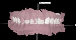
The mock-up of the future teeth was 3D printed upon completion of the modeling. A silicone key was obtained from the obtained model, by means of which the shape of the future teeth was transferred to the vestibular surface of the patient's anterior group of teeth using the Luxatemp light-cured composite dental material (Fig. 4).
Conclusion
The proposed method makes it possible to identically reproduce the results of a 2D smile design in a 3D treatment scene and to make a prototype of future restorations. It is already possible to create a 2D design within a few minutes during the initial consultation. This will be identically reproduced in the form of try-in restorations for coordination with the patient. With the Avantis 3D software, the 2D design stage can be used not only as a motivational effect, but also as a full-fledged element of complex digital planning of a dental patient.
References
- Coachman C., Calamita M.A., Sesma N. Dynamic Documentation of the Smile and the 2D/3D Digital Smile Design Process. Int J Periodontics Restorative Dent. 2017; 37 (2): 1831—93. PMID: 28196157
- Apresyan S.V., Stepanov A.G., Vardanyan B.A. Digital protocol for comprehensive planning of dental treatment. Clinical case analysis. Stomatology. 2021; 100 (3): 65—71 (In Russ.). eLIBRARY ID: 46222733
- Apresyan S.V., Suonio V.K., Stepanov A.G., Kovalskaya T.V. Evaluation of functional potential of CAD-programs in integrated digital planning of dental treatment. Russian Journal of Dentistry. 2020; 24 (3): 1311—34 (In Russ.). eLIBRARY ID: 44005657
- Apresyan S.V., Stepanov A.G., Retinskaya M.V., Suonio V.K. Development of complex of digital planning of dental treatment and assessment of its clinical effectiveness. Russian Journal of Dentistry. 2020;2 4 (3): 1351—40 (In Russ.). eLIBRARY ID: 44005658
- Ryahovsky A.N. 3D analysis of occlusal surfaces of teeth and their contacts. Part I. Development of a method for assessing the area of the occlusal surface, the severity of its relief and the histogram of contacts. Stomatology. 2021; 100 (4): 374—3 (In Russ.). eLIBRARY ID: 46390873
- Ryakhovsky A.N. A new concept of virtual 4D planning in dentistry. Digital dentistry. 2019; 10 (1): 112—1 (In Russ.). eLIBRARY ID: 39165059
- Zimmermann M., Mehl A. Virtual smile design systems: a current review. Int J Comput Dent. 2015; 18 (4): 3031—7. PMID: 26734665


