DOI:
10.37988/1811-153X_2021_4_56The significance of introducing ergonomic working techniques in the carpal tunnel syndrome prevention in dentists when performing local anesthesia
Downloads
Abstract
The aim was to assess the anatomo-functional changes in the carpal tunnel lumen during standard and ergonomic methodology of holding the carpulo-injector in dentistry.Materials and methods.
100 dentists aged 30 to 45 years (average 38±6 years) without carpal tunnel pathology were examined. As part of the experiment, conduction (mandibular) anesthesia was carried out on a biological material (head) under conditions close to natural, using a disposable carpool syringe-injector. Comparative analysis of carpal tunnel lumen was carried out according to the ultrasound examination data.
Results.
Under the the standard technique of holding the syringe-injector, a significant change in all parameters of the carpal tunnel lumen were noted: while its width was considerably reduced, its maximum height was increased in parallel due to wrist flexor tendon strain, which may contribute to carpal tunnel syndrome. Under the ergonomic technique of holding the syringe-injector, the anatomo-functional parameters of the carpal tunnel lumen did not significantly change, which indicates less tension load on the median nerve from the tendons of flexor muscles.
Conclusion.
The results of the study showed that ergonomic methods of carpula syringe holding during perduction anaesthesia can reduce the influence of tension load on the median nerve from the flexor muscles’ tendons, which will help prevent the carpal tunnel syndrome in dentists.
Key words:
dentist, tunnel carpal syndrome, ergonomics, carpel syringe-injector, honest sign, local anesthesiaFor Citation
Introduction
Compression-ischemic neuropathy of the median nerve, better known as carpal tunnel syndrome (CTS), is defined as a common pathology of the peripheral nervous system, affecting 3 to 6% of the population according to the literature [1], with a frequency of 150 per 100,000 people [2]. In recent years, there is evidence that the prevalence of this pathology is significantly higher in individuals whose activities involve prolonged and repeated flexion-extension hand movements [3]. The range of these occupations includes office workers, graphic designers, computer programmers, musicians, hairdressers, cashiers, and drivers [4, 5]. In healthcare, the risk group for the CTS development is primarily represented by dentists [6—10]. Unfortunately, in recent years, there is a negative trend in the CTS cases' increase among specialists in this medical field, as evidenced by the increasing professional interest in this problem and the growing number of scientific articles associated with this nosology (Fig. 1).
Thus, numerous studies established that almost every third dentist (30.5—36.5%) has characteristic manifestations of this disease [1, 11, 12], primarily sensory (numbness, paresthesias and pain) and motor (weakness, increased fatigue and reduced function) disorders [13]. Other studies indicate that signs of compression mononeuropathy of varying severity were observed in 86.0% of practitioners in this specialty [14]. The identified symptoms are more often observed in female dentists, and their prevalence increases significantly with age and work experience [1, 11, 15—17]. It was also shown that long work periods and more patients per day significantly increased the CTS risk [12, 16].
The high CTS prevalence among dentists indicates the advisability of studying the risk factors of the professional environment and implementing ergonomic work principles to prevent this pathology occurrence [11, 12, 15]. One of the most common dental treatment manipulations that create prolonged tension in the physician's wrist muscles is local anesthesia, as it is characterized by the need for slow drug administration (no more than ml/min) and is performed on weight, without supporting the patient [18, 19] In this regard, it seems relevant to substantiate the predominant use of ergonomic techniques in this procedure over traditional techniques, based on the control of carpal tunnel volume.
The aim of the study was to assess the anatomico-functional changes in the carpal tunnel lumen during standard and ergonomic carpulo-injector retention techniques in dentistry.
Materials and methods
The study included 100 dentists aged 30 to 45 years (average — 38.38±6.02 years) without carpal tunnel pathology.
Inclusion criteria: continuous medical experience, experience in using carpool technology of local anesthesia (reusable and disposable), individual voluntary consent to participate in the work, no contraindications to ultrasound examination. Exclusion criteria: peripheral nervous system disorders, hand and forearm injuries.
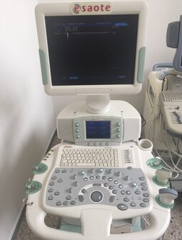
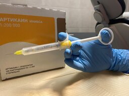
As part of the experiment, conduction (mandibular) anesthesia was performed on a biological material (head) under conditions close to natural, using a disposable carpool syringe-injector, a disposable carpool needle, and a disposable anesthetic. The carpal tunnel lumen was measured on an Esaote MyLaB 70 apparatus (Italy) with a 4—13 MHz linear transducer (Fig. 2). According to meta-analyzes, the high specificity and sensitivity of this method in the CTS diagnosis were proved [20—22].
The width and maximum height of the carpal tunnel was determined, visualized by ultrasound examination (US) between the wrist flexor retainer and the anterior edge of the proximal wrist bones. For clarity, the latter was divided into the height of the deep part of the carpal tunnel, whose dimensions were fixed from the tendons and synovial sheaths of the flexors to the wrist bones, and into the height of the wrist flexor tendons [23]. Wrist ultrasound was performed at rest, with the hand palm upward in passive flexion; in the working position under the standard syringe-injector holding technique and under the ergonomic instrument holding technique. In the standard technique, the thumb rests on the syringe-injector piston and the index and middle fingers hold the finger pad. Under the ergonomic technique, the thumb also rests on the syringe-injector piston, but the finger pad is held by index and ring fingers, while the middle finger compensates for the piston centralization and relieves hand tension [24].
Results
At rest, the carpal tunnel width was 23.43±0.45 mm, the height of its deep part was 1.9±0.1 mm, and the maximum height of the carpal tunnel was 9.4±0.89 mm.
Under the standard syringe-injector holding technique, a significant change in all parameters of the carpal tunnel lumen is noted: its width was significantly reduced, while its maximal height was increased due to wrist flexor tendon strain, which may contribute to (see the table).
| Indicator (mm) | At rest | Working position under the standard syringe-injector holding technique | Working position under the ergonomic instrument holding technique |
| Carpal tunnel width | 23.40±0.45 | 21.10±0.03* | 22.00±0.12 |
| Tendon height | 7.5±0.1 | 8.6±0.1* | 8.0±0.2 |
| Deep part height | 1.9±0.1 | 1.5±0.3* | 2.0±0.1 |
| Maximum carpal tunnel height | 9.40±0.89 | 10.10±0.11* | 10.00±0.93 |
| * — the differences are significant in comparison with the indicators measured at rest (p<0.05). | |||
Under the ergonomic syringe-injector holding technique the anatomico-physiological parameters of the carpal tunnel lumen did not change significantly, indicating less stress load on the median nerve from the flexor tendons.
Discussion
The results of this study indicate that under the standard syringe-injector holding technique during local anesthesia, the prolonged forced position of the doctor's working hand, the predominant static load and the high resistance of the jaw tissue lead to a significant decrease in carpal tunnel height, which, with repeated over a long time may contribute to the CTS occurrence in dentists and be a serious risk factor for the musculoskeletal disorders, that, according to the literature, are associated with this profession [11—15].
Understanding the reasons for this compression mononeuropathy became the basis for the development of ergonomic techniques that reduce the load on the tendons of the wrist flexor muscles and prevent pronounced narrowing of the carpal tunnel lumen. The successful application of ergonomics guarantees increased productivity of specialists, prevention of occupational diseases and injuries [26]. Studies aimed at improving ergonomics indicate the need to unload the working hand [27—29].
Indeed, engaging the ring finger and using the freed middle finger to centralise the piston relieves the tension of the dentist's working hand and significantly neutralizes the decrease in the carpal tunnel lumen (Fig. 3). The use of disposable, lightweight syringes-injectors not only plays an important role in preventing infectious diseases in patients, but also reduces the load on the dentist's hand muscles, preventing the CTS occurrence.
Conclusions
The results of the study showed that performing local anesthesia according to standard technique is accompanied by a significant decrease in the carpal tunnel lumen. It was proved that using ergonomic techniques for holding the carpule syringe-injector during this procedure reduces the influence of tension load on the median nerve from the flexor muscle tendons, which will help prevent the CTS occurrence in dentists. The developed ergonomic technique should be recommended in the educational process as a new technique for holding the carpool syringe during local anesthesia in the maxillofacial region. The use of anesthetics labeled by the national system “Honest Sing” will significantly reduce the amount of counterfeit, low-quality analogues and raise the level of Russians' safety.
According to paragraph 4032 of Russian Health Codes and Regulations 3.3686-21 (effective from September 1, 2021), there is an emphasis on using single-use carpel syringes, with their subsequent decontamination or disinfection as class B waste instead of reusable carpel syringes, as there is a high risk of needle injury staff when preparing them for sterilisation.
The use of disposable lightweight syringes-injectors not only plays an important role in the prevention of infectious diseases, both in medical staff in case of hand injuries during syringe preparation for sterilization and for patients, but also reduces the load on the dentist's hand muscles, preventing the CTS occurrence. This study showed the need to introduce the ergonomic principles in into the educational dentistry process, which will be the prevention of occupational hand disease in dentists.
References
- Alhusain F.A., Almohrij M., Althukeir F., Alshater A., Alghamdi B., Masuadi E., Basudan A. Prevalence of carpal tunnel syndrome symptoms among dentists working in Riyadh. Ann Saudi Med. 2019; 39 (2): 104—111. PMID: 30905925
- Yusupova D.G., Suponeva N.A., Zimin A.A., Zaytsev A.B., Belova N.V., Chechotkin A.O., Gushcha A.O., Gatina G.A., Polekhina N.V., Bundhun P., Ashrafov V.M. Validation of the Boston Carpal Tunnel Questionnaire in Russia. Neuromuscular Diseases. 2018; 8(1): 38—45 (In Russ.). eLIBRARY ID: 32850536
- Sevy J.O., Varacallo M. Carpal tunnel syndrome. Treasure Island (FL): StatPearls. 2021. PMID: 28846321
- Coraci D., Bellavia M.A., Hobson-Webb L., Santilli V., Padua L. Carpal tunnel syndrome and its relationship with occupation and sex: from objective evaluation to patients’ care. Occup Environ Med. 2017; 74 (2): 155. PMID: 27879351
- Lee I.H., Kim Y.K., Kang D.M., Kim S.Y., Kim I.A., Kim E.M. Distribution of age, gender, and occupation among individuals with carpal tunnel syndrome based on the National Health Insurance data and National Employment Insurance data. Ann Occup Environ Med. 2019; 31: e31. PMID: 31737286
- de Krom M.C., de Krom C.J., Spaans F. [Carpal tunnel syndrome: diagnosis, treatment, prevention and its relevance to dentistry]. Ned Tijdschr Tandheelkd. 2009; 116 (2): 97—101 (In Dutch). PMID: 19280893
- Sakzewski L., Naser-ud-Din S. Work-related musculoskeletal disorders in Australian dentists and orthodontists: Risk assessment and prevention. Work. 2015; 52 (3): 559—79. PMID: 26409367
- de Jesus Júnior L.C., Tedesco T.K., Macedo M.C., Agra C.M., Mello-Moura A.C., Morimoto S. A self-report joint damage and musculoskeletal disorders data among dentists: a cross-sectional study. Minerva Stomatol. 2018; 67 (2): 62—67. PMID: 29446269
- Jaoude S.B., Naaman N., Nehme E., Gebeily J., Daou M. Work-Related musculoskeletal pain among lebanese dentists: An epidemiological study. Niger J Clin Pract. 2017; 20 (8): 1002—1009. PMID: 28891546
- Occhionero V., Korpinen L., Gobba F. Upper limb musculoskeletal disorders in healthcare personnel. Ergonomics. 2014; 57 (8): 1166—91. PMID: 24840049
- Harris M.L., Sentner S.M., Doucette H.J., Brillant M.G.S. Musculoskeletal disorders among dental hygienists in Canada. Can J Dent Hyg. 2020; 54 (2): 61—67. PMID: 33240365
- Gandolfi M.G., Zamparini F., Spinelli A., Risi A., Prati C. Musculoskeletal Disorders among Italian Dentists and Dental Hygienists. Int J Environ Res Public Health. 2021; 18 (5): 2705. PMID: 33800193
- Abichandani S., Shaikh S., Nadiger R. Carpal tunnel syndrome an occupational hazard facing dentistry. Int Dent J. 2013; 63 (5): 230—6. PMID: 24074016
- Prasad D.A., Appachu D., Kamath V., Prasad D.K. Prevalence of low back pain and carpal tunnel syndrome among dental practitioners in Dakshina Kannada and Coorg District. Indian J Dent Res. 2017; 28 (2): 126—132. PMID: 28611320
- Meisha D.E., Alsharqawi N.S., Samarah A.A., Al-Ghamdi M.Y. Prevalence of work-related musculoskeletal disorders and ergonomic practice among dentists in Jeddah, Saudi Arabia. Clin Cosmet Investig Dent. 2019; 11: 171—179. PMID: 31308760
- Borhan Haghighi A., Khosropanah H., Vahidnia F., Esmailzadeh S., Emami Z. Association of dental practice as a risk factor in the development of carpal tunnel syndrome. J Dent (Shiraz). 2013; 14 (1): 37—40. PMID: 24724115
- Maghsoudipour M., Hosseini F., Coh P., Garib S. Evaluation of occupational and non-occupational risk factors associated with carpal tunnel syndrome in dentists. Work. 2021; 69 (1): 181—186. PMID: 33998581
- Vasil’ev Y.L., Rabinovich S.A., Dydykin S.S., Bogoyavlenskaya T.A., Kashtanov A.D., Kuznetsov A.I. Evaluation of dentists regulatory systems stress during the provision of dental care according to pulse oximetry data. Stomatology. 2020; 99(6): 89—93 (In Russ.). eLIBRARY ID: 44298780
- Nowak J., Erbe C., Hauck I., Groneberg D.A., Hermanns I., Ellegast R., Ditchen D., Ohlendorf D. Motion analysis in the field of dentistry: a kinematic comparison of dentists and orthodontists. BMJ Open. 2016; 6 (8): e011559. PMID: 27531728
- Erickson M., Lawrence M., Lucado A. The role of diagnostic ultrasound in the examination of carpal tunnel syndrome: an update and systematic review. J Hand Ther. 2021; S0894-1130(21)00061-2. PMID: 34261588
- Horng M.H., Yang C.W., Sun Y.N., Yang T.H. DeepNerve: A New Convolutional Neural Network for the Localization and Segmentation of the Median Nerve in Ultrasound Image Sequences. Ultrasound Med Biol. 2020; 46 (9): 2439—2452. PMID: 32527593
- Fowler J.R., Gaughan J.P., Ilyas A.M. The sensitivity and specificity of ultrasound for the diagnosis of carpal tunnel syndrome: a meta-analysis. Clin Orthop Relat Res. 2011; 469 (4): 1089—94. PMID: 20963527
- Bianchi S., Beaulieu J.Y., Poletti P.A. Ultrasound of the ulnar-palmar region of the wrist: normal anatomy and anatomic variations. J Ultrasound. 2020; 23 (3): 365—378. PMID: 32385814
- Vasil’ev Y, Meylanova R., Rabinovich S. Assessment of the motor function of the hand in dentists with subclinical manifestations of carpal syndrome with local anesthesia. Russian Journal of Pain. 2017; 3—4 (54): 54—9 (In Russ.). eLIBRARY ID: 32724442
- Haghighat A., Khosrawi S., Kelishadi A., Sajadieh S., Badrian H. Prevalence of clinical findings of carpal tunnel syndrome in Isfahanian dentists. Adv Biomed Res. 2012; 1: 13. PMID: 23210072
- Gupta A., Bhat M., Mohammed T., Bansal N., Gupta G. Ergonomics in dentistry. Int J Clin Pediatr Dent. 2014; 7 (1): 30—4. PMID: 25206234
- De Sio S., Traversini V., Rinaldo F., Colasanti V., Buomprisco G., Perri R., Mormone F., La Torre G., Guerra F. Ergonomic risk and preventive measures of musculoskeletal disorders in the dentistry environment: an umbrella review. PeerJ. 2018; 6: e4154. PMID: 29362689
- Vasil’ev Yu.L., Ivanov A.A., Smilyk I.M., Kashtanov A.D., Kytko O.V. Dissection courses as a way to change the paradigm of modern comprehensive dental education. Clinical Dentistry (Russia). 2021; 24 (2): 130—4 (In Russ.). eLIBRARY ID: 46322613
- Rabinovich S.A., Vasil’ev Yu.L. Modern injector systems in dentistry. Part 2: use of one-time syringes in aspect of individual approach. Clinical Dentistry (Russia). 2014; 2 (70): 26—9 (In Russ.). eLIBRARY ID: 23797666


