Difficulty and strategy of treatment for dental patients with epilepsia
Downloads
Abstract
Retrospective analysis of patients with epilepsia during splint-therapy treatment. Patients suffered from pain in temporomandibular and masticate muscles areas.Methods.
Visual analisis, splint-therapy (splint), gnatodynamometry, superficial electromyography.
Result.
Concern among patients with epilepsia splint-therapy is able to adapt temporo-mandibular and masticate muscles for adequate function, avoid pain syndrome, and lead to more successful prosthetic dental treatment. Level of points in gnatodynamometry increased after 3-months splint-therapy treatment compared to level of points in superficial electromyography, where changes was insignificant. Pain symptomathy has been significantly decreased by objectively observed of patients; based on the opinions from themselves.
Conclusion.
Patients with epilepsia necessary needed for wear a hard acrylic splint on mandible to all night long and 2—3 hours per day plus, and also visit prosthodontist for dynamic control and corrections not on less than 1 per 3 months.
Key words:
epilepsia, teeth abrasion, tooth crown, splint-therapy, splintFor Citation
Introduction
Patients with epilepsia presented the particular category among dental patients. It deals with possible provocation of status epilepticus in dental clinic or hospital by special attractors (bright light; loud noise; unexpected touching). According to international classification there are several types of epilepsia: generalized (primary total activation in nervous cells of brain) and focal (activation in separate group of some neurones) [1].
Concern to etiology, epilepsia include of idiopathic form (unknown etiology); symptomatic form (as a single symptom of other disease); genetic form [1, 2]. Beside that, there are 3 separate categories: progressive myoclonical epilepsy; encephalophaties from epilepsia; reflex epilepsies (idiopathic photosensitive occipital epilepsia that might be provocated by bright light; startle-epilepsia, which connected with loud, high-frequency noise, unexpected startle) [1, 2]. There are some special forms for every level of age. Type of treatment concern of age of epilepsia debut and form of manifestation (tonical, psycho-motorical, tonic-clonic form: grand mal — most difficult form). The focal reason of epilepsia pathogenesis provided by synchronized impulses in group of neurones towards to neurone cell membrane injury and disbalance between upload and down load polarized membrane's potential. It connected with influence of ion-target neurotransmitters: 4-aminobutanoic acid increased penetration of neurone cell membrane for chlor anions inside cell; N-methyl-D-aspartate have decreased penetration for natrium and calcium cations. Result of that is an effect of depolarization in intercellular stretch (increase of postsynaptic potential) [1].
Planning of most traumatic and long dental procedures (extraction of third molar teeth; reconstructive plastic surgery) necessary to provide in dental hospital with assistance of anesthesiologists and resuscitologists. It deals with possibility appearance of status epilepticus (long, often, exaggerated type of epilepsia manifestation) [1, 3, 4]. Duration of manifestation connect with decrease of 4-aminobutanoic acid and increase of N-methyl-D-aspartate influence, and afterwards rise up of calcium cations in liquor, that lead to tonic clonic muscle movements. It can be cause of aspiration pneumonia, also head and jaws injures, fractures of teeth [1, 4, 5].
Aim of study: Improve prosthetic dental supervision among patients with epilepsia.
Materials and methods
During the period of time from 2017 to 2019 — 7 patients with epilepsia in age of 20—45 years old was examined. They suffered from night-time bruxism; non of deal with epilepsia manifestations. Patients marked pain symptoms in area of ears and area of m.masseter from both sides. Bruxism was compilated with pathologic movements of single molars, premolars. Palpation appeared absence of masticatory muscles hypertrophia. There was multiple fractures of teeth; hard fractured teeth was extracted initially. And afterwards the solid acrylic mouthguards on low jaw (splints) was completed for all patients. Splints was necessary to use in whole night-time period and about 2—3 hours during the day. Dynamic control for patients appeared from 3-months period [6—8].
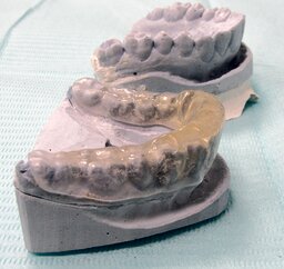
For registration masticate force (force of bite) and level of periodont resistance gnatodynamometry was used. If level of strength in bite of frontal teeth was under 50 N (Newton) it was diagnosed as temporo-mandibular joint disfunction. Throughout procedure it's needed for patients to be relaxed with some kind of comfortable feeling. Gnatodynamometry was examined preliminary and 3 months after treatment. According to this superficial electromyography at m.masseter area was used too. The first electrode was localized on border between upper and middle part from angle of mandible to foramen auricularis. The second electrode was located beside and lower the first one. It was 5-minute recording from both sides. Afterwards, the amplitude of tone (spontaneous) activity of impulse in rest position was verified. Examination was also before and after 3 months of splint-therapy (solid acrylic mouthguard on lower jaw, Fig. 1) [9—13].
Due to period of 3 months — capacity for structural, stabilized changes of muscle adaptation will appeared. This dynamic biochemical aspects concern with muscle metabolism, increase of numbers of mitochondria in muscle cells and ATP (adenosine triphosphate) production [14, 15].
Results
After 3 months mouthguard treatment the level of points in gnatodynamometry in frontal teeth verified increased level compared to level at the beginning (33.9±2.4; 45.1±1.8 N). The significant distinction in numbers of superficial electromyography was not indicated. Intuitively, the patients marked evaporation of headache and ache symptoms in locale of ears.
Conclusion
G.N. Krijanovskiy marked, that rise up the points of pathological system lead to their more higher level of resistance compared to complex contain from just 2 points. It appears in epileptic complex very clearly. Range of resistance inside pathological system able to turn out to be higher level of resistance towards to their long-term manifestation. The particular kind of pathological system concerned with licks of their parts that might be more exaggerate. Therefore, for result of pathological system to be erasure — the blocking of primary point could be not adequate [16, 17].
The structural organization of pathological and physiological (functional) system are distinguished actually. By terms of P.K. Anohin (1973), the functional system contains of dynamically mobilized structures in whole. The functional principle of separate mobilisation of structures is able to be dominated. In any case, without exception, physiological system is actual functional system [18, 19].
According to principles of A.M. Vein (1998), that concerned with psychoneurological reactions: the healthy human is often able to reacted separately; it determined by psychophysiological pattern; and in case of illness — it determined by diffusal, non control activation of all psychovegetative patterns. It could be as well on higher levels (cerebral vegetative reactions) as on lower levels (facial and trigeminal nervous activity, for pattern teeth grinding) [20—25]. The better level of points in gnatodynamometry might inform about some sort of local normalization in temporo-mandibular joints and partly in masticatory muscles. The absence of significant verification in numbers of superficial electromyography apparently due to impossibility of blocking the primarily dominant points in subcortical structures and in trigger-points of masticate muscles [26—28].
Recommendation for pharmacological therapy (antiepilept. Topiramat in rus. pharm.) should be proscribed by prescription very carefully. The prosthodontic splint (solid acrylic mouthguard) must to be covering for all teeth and alveolar parts absolutely. Using the face bow and registers of bite in central occlusion is needed to be for sure [29—32].
Dental patients with epilepsia also are needed to provide examination with control of splint condition (appearance of some balance, defects) — 1 time for each 3 months; the cone beam computed tomography and MRI ought to be presented: for control of treatment [33—37].
References
- Braun R.T., Holmes G.L. Epilepsy. Clinical guidance. Moscow: Binom, 2014. Pp. 75—180 (In Russ.).
- Engel J. jr, International League Against Epilepsy (ILAE). A proposed diagnostic scheme for people with epileptic seizures and with epilepsy: report of the ILAE Task Force on Classification and Terminology. Epilepsia. 2001; 42 (6): 796—803. PMID: 11422340.
- DeLorenzo R.J., Pellock J.M., Towne A.R., Boggs J.G. Epidemiology of status epilepticus. J Clin Neurophysiol. 1995; 12 (4): 316—25. PMID: 7560020.
- DeLorenzo R.J., Waterhouse E.J., Towne A.R., Boggs J.G., Ko D., DeLorenzo G.A., Brown A., Garnett L. Persistent nonconvulsive status epilepticus after the control of convulsive status epilepticus. Epilepsia. 1998; 39 (8): 833—40. PMID: 9701373.
- Drislane F.W. Evidence against permanent neurologic damage from nonconvulsive status epilepticus. J Clin Neurophysiol. 1999; 16 (4): 323—31. PMID: 10478705.
- Sisolyatin N.G. Classification of temporo-mandibular joint diseases. Stomatology. 1999; 78 (2): 34—9 (In Russ.).
- Sisolyatin N.G. Actual questions in diagnostic and treatment of temporo-mandibular joints injures. Stomatology. 1999; 78 (2): 34—9 (In Russ.).
- Sisolyatin N.G. Classification of temporo-mandibular joints diseases and injures. Moscow: Medical Book, 2001. Pp. 70—78 (In Russ.).
- Hvatova V.A. Temporo-mandibular joints diseases and methods of treatment. Part 2. Functional anatomy and biomechanic in temporo-mandibular joint. New in dentistry. 1997; 8: 22—24 (In Russ.).
- Hvatova V.A. Clinical gnathology. Moscow: Medicine, 2011. 296 p. (In Russ.).
- Minyaeva V.A. Problems of prosthetic dentures in madibular-facial area. Moscow: PoliMediaPress, 2007. 189 p. (In Russ.).
- Sergeeva T.A. Diagnostic and treatment of temporo-mandibular joints dysfunction: master‘s thesis abstract. St.Petersburg, 1997. 23 p. (In Russ.).
- Turbina L.G. Diagnostic and pathogenetic treatment in case of myofacial pain syndrome of face. Russian Journal of Dentistry. 2001; 5: 35—7 (In Russ.).
- Meerson F.Z. Pathogenesis and previous therapy of stress and ishemical heart injures. Moscow: Medicine, 1984. 269 p. (In Russ.).
- Meerson F.Z. Adaptation to stressful situations and physical exertion. Moscow: Medicine: 1988. 253 p. (In Russ.).
- Krijanovskiy G.N. Determinant structures in pathology of nervous system. Genenerator mechanisms of neuropathological syndromes. Moscow: Medicine, 1980. 358 p. (In Russ.).
- Krijanovskiy G.N. System mechanisms of nervous and psychiatric deviations. Common questions of neurology and psychiatry. 1996; 6: 5—11 (In Russ.).
- Anohin P.K. Principal questions in common theory of functional systems. Moscow: Science, 1973. Pp. 5—61 (In Russ.).
- Anohin P.K. A studies concern with physiology of functional systems. Moscow.: Medicine, 1975. 447 p. (In Russ.).
- Vein A.M. Vegetative disorders: Clinic, treatment, diagnostic. Moscow: MIA, 1998. Pp. 22—38 (In Russ.).
- Rubinov I.S. Physiological basics of dentistry. Leningrad: Medicine, 1965. 351 p. (In Russ.).
- Selye G. Studies (essays of adaptation syndrome. Moscow.: Medgiz, 1960. 254 p.) (In Russ.).
- Pusin M.N. Affective disorders in structure concern by diagnostic and treatment of temporo-mandibular joint dysfunction. Russian Journal of Dentistry. 2002; 5: 37—42 (In Russ.).
- Pusin M.N. Pain dysfunction of temporo-mandibular joint. Russian Journal of Dentistry. 2002; 1: 31—6 (In Russ.).
- Pusin M.N. Diagnostic and treatment of pain dysfunction of temporo-mandibular joint in conditions of special neuro-stomatological cabinet. Russian Journal of Dentistry. 2002; 2: 28—30 (In Russ.).
- Pusin M.N. Pain dysfunction of temporo-mandibular joint. Moscow: Medicine, 2002. 158 p. (In Russ.).
- Semenov I.J. Neuro-gumoral aspects of syndrome temporo-mandibular pain joint dysfunction: master‘s thesis abstract. Moscow, 1997. 18 p. (In Russ.).
- Semkin V.A. Clinical-radiological appearances of muscles dysbalance concern with temporo-mandibular joint and its treatment. Stomatology. 1997; 76 (5): 15—7 (In Russ.).
- Badanin V.V. Diagnostic and prosthetic treatment in case of temporo-mandibular joints diseases. International Dental Review.2000; 2: 8—12 (In Russ.).
- Badanin V.V. Occlusion disorder the main etiological factor in temporo-mandibular joints dysfunction. Stomatology. 2000; 79 (1): 51—4 (In Russ.).
- Badanin V.V. Occlusal splints effective method in prosthetic treatment of functional disorders of temporo-mandibular joint. The Dental Institute. 2003; 3: 26—30 (In Russ.).
- Kato T., Lavigne G.J. Sleep bruxism: A sleep-related movement disorder. Sleep Medicine Clinics. 2010; 5 (1): 9—35. DOI: 10.1016/j.jsmc.2009.09.003.
- Wohlberg V., Schwahn C., Gesch D., Meyer G., Kocher T., Bernhardt O. The association between anterior crossbite, deep bite and temporomandibular joint morphology validated by magnetic resonance imaging in an adult non-patient group. Ann Anat. 2012; 194 (4): 339—44. PMID: 21646004.
- Wu C.-K., Hsu J.-T., Shen Y.-W., Chen J.-H., Shen W.-C., Fuh L.-J. Assessments of inclinations of the mandibular fossa by computed tomography in an Asian population. Clin Oral Investig. 2012; 16 (2): 443—50. PMID: 21318300.
- Vojdani M., Bahrani F., Ghadiri P. The study of relationship between reported temporomandibular symptoms and clinical dysfunction index among university students in Shiraz. Dent Res J (Isfahan). 2012; 9 (2): 221—5. PMID: 22623942.
- Türp J.C., Schindler H. The dental occlusion as a suspected cause for TMDs: epidemiological and etiological considerations. J Oral Rehabil. 2012; 39 (7): 502—12. PMID: 22486535.
- Sümbüllü M.A., Cağlayan F., Akgül H.M., Yilmaz A.B. Radiological examination of the articular eminence morphology using cone beam CT. Dentomaxillofac Radiol. 2012; 41 (3): 234—40. PMID: 22074873.


