Evaluation of an integrated approach to the restoration of the occlusal surface of the coronal part of the tooth using computer technology
Downloads
Abstract
The aim of the study was to evaluate the long-term results of the restoration of posterior teeth using the developed computerized technique for restoring the occlusal surface, taking into account the individual morphometric parameters of the patient’s dental crown. Objectives: 1) to develop and test, using various composite materials, an original method of aesthetic and functional restoration of hard tissues of teeth in young people with the stage of computer calculation of the parameters of the occlusal surface, taking into account the identified individual morphometric parameters; 2) to evaluate in a comparative aspect the long-term results of the application of the methodology of restorative dental treatment developed on the basis of computer technologies.Materials and methods.
The objects of the study were: caries-resistant patients (82 people aged 18 to 35 years, male and female) and 106 patients, of different sex, who had carious lesions and complications of caries (pulpitis and periodontitis).
Results.
On the basis of the data obtained, an innovative program was developed (certificate of state registration of the computer program No. 2018611780 dated 02/07/2018) and introduced an innovative program for calculating the dimensional characteristics of the occlusal surface of the teeth. At the end of the treatment carried out using the author’s and traditional methods of restoration of teeth, there is a positive dynamics of clinical indicators of the state of organs and tissues of the mouth.
Conclusion.
The created innovative program for calculating the lost tissues of the occlusal surface of the tooth allows dentists to reconstruct the hard tissues of the teeth, taking into account the individual dimensional characteristics of the patient’s dentition, which contributes to improving the quality of dental care to the population.
Key words:
restoration, computer program, aesthetic dentistryFor Citation
Introduction
An analysis of the degree of development of the problem indicates that modern conservative dentistry has a wide range of composite materials for the aesthetic and functional restoration of lateral teeth [1—6]. Traditional methods of dental restoration are constantly being improved and new methods of restorative treatment of lost hard tissues of teeth are being created [7—13]. Despite the active introduction of these effective techniques into everyday practice, the quality and stability of the results obtained do not always meet the patient's subjective requirements and the doctor's objective conclusions [14—21].
The development of innovative computer programs to optimize modern methods of dental restoration and their implementation at a therapeutic appointment can provide additional important information for a comprehensive individualized assessment of the patient's dental status. This, in turn, can help in choosing an individual plan for restorative dental treatment in a particular patient, which will allow for the reconstruction of the tooth crown, taking into account its smallest details, such as the occlusal surface, while ensuring optimal functioning of the entire dentoalveolar apparatus [22—29].
The aim of the study was to evaluate the long-term results of the performed restorations of the posterior teeth using the developed computerized technique for restoring the occlusal surface, taking into account the individual morphometric parameters of the patient's dental crowns. To achieve this goal, the following tasks have been set in the present study:
To develop and test, using various composite materials, an original method for the aesthetic and functional restoration of hard dental tissues.
To assess, in a comparative manner, the long-term results of the application of the methodology of restorative dental treatment developed on the basis of computer technologies.
Materials and methods
The theoretical and methodological basis of the study included inferences from scientific and practical conferences, the works of domestic and foreign scientists on the problems of tooth restoration using computer technology, and modern composite materials. The objects of the study were patients aged 18 to 35 years, belonging to different sexes, who were divided into two groups:
- 82 caries-resistant people;
- 106 patients who had carious lesions and complications of caries — pulpitis and periodontitis.
In group I, a statistical study was carried out that proved the presence of correlations (95%) between the morphometric parameters of the teeth, which makes it possible to obtain certain patterns in their structure. Based on the data obtained, the equations of paired regressions were compiled. The exact significance of the differences, as shares in %, was assessed by the Fisher method with the calculation of the φ index. The Student's t-test was used for error calculation and the assessment of the significance of the regression.
Based on the results, a computer program was developed (certificate of state registration of a computer program No. 2018611780 dated 02/07/2018). A protocol for working with the author's program has also been introduced.
To calculate the missing parameters, a dentist-therapist measures teeth that have a correlation, and enters the obtained data into the program. Based on the regression equations, a calculation is carried out, and the missing parameters of the occlusal surface of the teeth become known.
In group II, an assessment according to Ridge's criteria was used to objectively assess the restoration of the occlusal surface of the coronal part of the posterior teeth (Ryge, 1975). The clinical assessment of the restored teeth was analyzed according to the criteria of “anatomical shape”, “surface and color”, and “marginal integrity”, based on the estimates from Romeo, Sierra, Tango, Victor codes. The occlusal surfaces of the teeth were restored with light-cured composite materials, using various modeling techniques, in patients of the main group as well as the comparison group. Clinical evaluations were performed one week after patient debridement and were repeated after the first year and after two years.
Results and discussion
In group I, 162 models were obtained, on which 12,300 measurements were carried out (see the example of one of the patients in the table). A total of 1,312 teeth were examined. Dental patient records were entered for all the patients, after which all the obtained values were entered into a computer program. Based on the results of the stratification performed, 84,364 pairwise regression equations were compiled.
| Upper jaw | Length (mm) | Height (mm) | Width (mm) | Length (mm) | Height (mm) | Width (mm) |
| Tooth 1.6 | Tooth 2.6 | |||||
| Medial buccal tubercle | 5.8 | 6.8 | 6.0 | 5.6 | 6.9 | 6.0 |
| Distal buccal tubercle | 4.6 | 6.4 | 5.7 | 4.6 | 6.5 | 5.8 |
| Medial palatal tubercle | 6.0 | 7.0 | 6,7 | 5.8 | 6.9 | 6.8 |
| Distal palatal tubercle | 4.0 | 5.5 | 4.0 | 3.9 | 5.4 | 3.9 |
| Lower jaw | Tooth 4.6 | Tooth 3.6 | ||||
| Medial buccal tubercle | 4.8 | 6.3 | 4.5 | 4.8 | 6.2 | 4.5 |
| Median tubercle | 4.28 | 6 | 3.4 | 4.25 | 6 | 3.5 |
| Distal buccal tubercle | 4.0 | 5.8 | 2.6 | 3.5 | 5.7 | 2.5 |
| Medial palatal tubercle | 4.5 | 6.5 | 5.7 | 4.6 | 6.5 | 5.8 |
| Distal palatal tubercle | 4.5 | 6.3 | 5.1 | 3.5 | 6.2 | 5.0 |
In group II, a comparison was made between the author's method and the traditional methods of restoration. For this purpose, the patients of this group were further divided into the main subgroup, where the restoration of teeth was carried out using the author's method, and the subgroup of comparison, in which layer-by-layer modeling, the traditional method of restoration of teeth, was used.
Based on the data obtained after an assessment of 421 fillings in terms of the quality of the location of the restoration, it was observed that in the cases where the author's (223 fillings) method was applied, the results were better in a year (97%) and two years later (91%). With the traditional restoration technique (198 fillings), the quality of the restoration location was 78% and 67% after one year and two years, respectively. The quality of processing, final polishing, and color rendering of the restored fillings also positively differ. The preservation of the relief and shape of the filling as a whole and the absence of coloration of the border of the “tooth-to-composite” transition were also assessed. The use of an innovative restoration technique (223 fillings) made it possible to obtain the best result after a year (95%) and after two years (85%), in comparison with the results of restoration using the traditional (198 fillings) technique after one year (76%) and after two years (63%). When assessing the quality of the marginal fit of fillings/tooth restoration, the use of the author's technique (223 fillings) made it possible to obtain the best result after a year (91%) and after two years (80%), in contrast to the traditional method (198 fillings), in which 71% cases were reported after a year and 47% after two years.
Clinical case
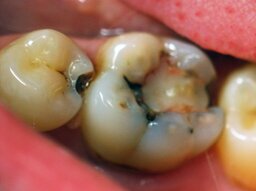
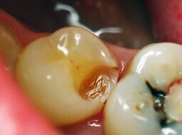
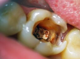
A clinical case of restoration of the coronal part of teeth 35 and 36 using the author's computer program is demonstrated. The restoration of the destroyed coronal part was performed by a direct method in the mouth using a submicron universal composite material. Tooth 36 was previously treated for pulpitis, it has filling on the chewing surface, and its marginal fit is broken. The tooth also has a carious cavity on the distal contact surface (Tooth Occlusal Surface Failure Index by Milikevich about 0.3). The probing and percussion is painless, and on the roentgenogram the root canals are filled up to the apex. Tooth 35 has a medium-sized carious cavity on the distal contact surface. Probing is painful along the enamel-dentinal junction, while percussion is painless (Fig. 1). Diagnosis: Dentin caries of tooth 35 (K02.01), chronic periodontitis of tooth 36 (K04.03) (filling defect).
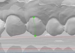
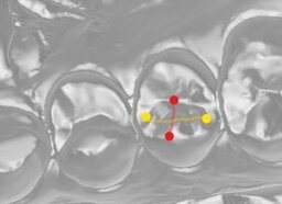
To obtain the missing parameters, the dentist-therapist, after intraoral scanning, measures the teeth with a correlation (Fig. 2, 3) and enters the obtained data into the developed computer program (Fig. 4). After that, with the help of a computer program, the available values are processed. On the basis of the regression equations, the calculation is carried out, and the missing values of the occlusal surface of the teeth become known (Fig. 5, 6).
Under conduction anesthesia, preparation of carious cavities and drug treatment were performed (Fig. 7, 8). With the help of the computer program, restoration of the dimensional characteristics of the occlusal surface of the tooth was carried out using a light-curing composite material. Final view of the reconstructed second premolar and first mandibular molar is shown in Fig. 9.
At the end of the treatment using the author's method and the traditional methods of restoration of teeth, positive dynamics of clinical indicators of the state of organs and tissues of the mouth was observed.
Conclusions
Thus, the program created by the author for calculating the lost tissues of the occlusal surface of the tooth allows dentists to reconstruct the hard tissues of the teeth, taking into account the individual dimensional characteristics of the patient's dentition, which helps to improve the quality of dental care in the population.
References
- Shatrov I.M., Zholudev S.E. Optimization of occlusal surface modeling for full ceramic restorations. Actual Problems in Dentistry. 2013; 1: 47—50 (In Russ.). eLIBRARY ID: 18976557
- Gilmiyarov E.M., Arnautov B.P. The quality of life of patients with caries of contact area on posterior teeth treated with different matrix systems. Izvestia of Samara Scientific Center of the Russian Academy of Sciences. 2015; 17 (2—2): 288—291 (In Russ.). eLIBRARY ID: 24118290
- Dutova A.O., Bryanskaya M.N., Shapovalov A.G. Modern technology of restoration of teeth with composite materials. Transbaikal Medical Journal. 2012; 2: 26—31 (In Russ.). eLIBRARY ID: 32565094
- Cervino G., Fiorillo L., Arzukanyan A.V., Spagnuolo G., Cicciù M. Dental restorative digital workflow: Digital smile design from aesthetic to function. Dent J (Basel). 2019; 7 (2): 30. PMID: 30925698
- Martins A.V., Albuquerque R.C., Santos T.R., Silveira L.M., Silveira R.R., Silva G.C., Silva N.R.F.A. Esthetic planning with a digital tool: A clinical report. J Prosthet Dent. 2017; 118 (6): 698—702. PMID: 28533014
- Naoum S.J., Ellakwa A., Morgan L., White K., Martin F.E., Lee I.B. Polymerization profile analysis of resin composite dental restorative materials in real time. J Dent. 2012; 40 (1): 64-70. PMID: 22044773
- Rybnikova E.P. Restoration of frontal units group. Clinical Dentistry (Russia). 2013; 4 (68): 76—81 (In Russ.). eLIBRARY ID: 22450783
- Lomiashvili L.M., Ayupova L.G., Pogadaev D.V., Mikhailovsky S.G. The art of modeling and restoration of teeth. 2nd edition. Omsk: Polygraph, 2014. 436 p. (In Russ.). eLIBRARY ID: 37751573
- Maksimova O.P. Anterior teeth aesthetics. Clinical dentistry (Russia). 2013; 3 (67): 20—23 (In Russ.). eLIBRARY ID: 22450760
- Rybnikova E.P. Anterior teeth restoration for rehabilitation of physiologic occlusal adjustment. Clinical Dentistry (Russia). 2012; 2 (62): 28—30 (In Russ.). eLIBRARY ID: 22473171
- Starodubova A.V., Vinnichenko Yu.A. Structural restoration of the crown part of the tooth with composite materials with the creation of a layer of artificial raincoat dentin. Russian Journal of Dentistry. 2018; 22 (3): 150—151 (In Russ.). eLIBRARY ID: 35419634
- Alsamadani K.H., Abdaziz el-S.M., Gad el-S. Influence of different restorative techniques on the strength of endodontically treated weakened roots. Int J Dent. 2012; 2012: 343712. PMID: 22666251
- Lowe R.A. No-prep veneers: a realistic option. Dent Today. 2010; 29 (5): 80—2, 84, 86. PMID: 20506914
- Stafeev A.A., Solov’ev S.I., Hizhuk A.V.1, Storozhenko V.Ju. Precision of chewing efficiency using a computer program “ChewingView”. Modern Prosthetic Dentistry. 2017; 28: 27—30 (In Russ.). eLIBRARY ID: 35312568
- Shylenko D.R., Gasanov R.A., Toncheva E.D., Skyrda L.Yu. Biomechanic analysisof the factors of the influencing longevity restorations of the masticatory group of teeth. World of Medicine and Biology. 2009; 2 (2): 72—77 (In Russ.). eLIBRARY ID: 22137953
- de Boer I.R., Bakker D.R., Wesselink P.R., Vervoorn J.M. [The Simodont in dental education]. Ned Tijdschr Tandheelkd. 2012; 119 (6): 294—300 (In Dutch). PMID: 22812267
- Ebert J., Frankenberger R., Petschelt A. A novel approach for filling tunnel-prepared teeth with composites of two different consistencies: a case presentation. Quintessence Int. 2012; 43 (2): 93—6. PMID: 22257869
- Omar D., Duarte C. The application of parameters for comprehensive smile esthetics by digital smile design programs: A review of literature. Saudi Dent J. 2018; 30 (1): 7—12. PMID: 30166865
- Suenaga H., Hoang Tran H., Liao H., Masamune K., Dohi T., Hoshi K., Mori Y., Takato T. Real-time in situ three-dimensional integral videography and surgical navigation using augmented reality: a pilot study. Int J Oral Sci. 2013; 5 (2): 98—102. PMID: 23703710
- Terry A.D., Geller W. An esthetic and restorative dentistry: material selection and technique. London: Quintessence, 2013. 752 p.
- Zanardi P.R., Zanardi R.L R., Stegun R.C., Sesma N., Costa B.N., Cruz Laganá D. The use of the digital smile design concept as an auxiliary tool in aesthetic rehabilitation: A Case Report. Open Dent J. 2016; 10: 28—34. PMID: 27006721
- Boldyrev Yu.A., Mandra Yu.V. Social significance of aesthetic and functional restoration of teeth by direct and indirect methods. Actual Problems in Dentistry. 2017; 4: 3—8 (In Russ.). eLIBRARY ID: 30638212
- Vedeneva E.V. The quality of life of patients seeking aesthetic dental care: master’s thesis. Moscow: Moscow State University of Medicine and Dentistry, 2010. 22 p. (In Russ.). eLIBRARY ID: 19318620
- Li R.W., Chow T.W., Matinlinna J.P. Ceramic dental biomaterials and CAD/CAM technology: state of the art. J Prosthodont Res. 2014; 58 (4): 208—16. PMID: 25172234
- Dietschi D., Argente A. A comprehensive and conservative approach for the restoration of abrasion and erosion. Part II: clinical procedures and case report. Eur J Esthet Dent. 2011; 6 (2): 142—59. PMID: 21734964
- Hollis W., Darnell L.A., Hottel T.L. Computer assisted learning: a new paradigm in dental education. J Tenn Dent Assoc. 2011; 91 (4): 14-8; quiz 18-9. PMID: 22256700
- Torabi K., Farjood E., Hamedani S. Rapid prototyping technologies and their applications in prosthodontics, a review of literature. J Dent (Shiraz). 2015; 16 (1): 1—9. PMID: 25759851
- Tassery H., Levallois B., Terrer E., Manton D.J., Otsuki M., Koubi S., Gugnani N., Panayotov I., Jacquot B., Cuisinier F., Rechmann P. Use of new minimum intervention dentistry technologies in caries management. Aust Dent J. 2013; 58 Suppl 1: 40—59. PMID: 23721337
- Vandenberghe B. The digital patient Imaging science in dentistry. J Dent. 2018; 74 Suppl 1: S21-S26. PMID: 29929585
