
DOI:
10.37988/1811-153X_2021_1_35Radiological characteristics of endodontic materials and fragments of broken instruments in the canals of extracted teeth according to computed tomography
Downloads
Abstract
Microfocus cone-beam computed tomography (micro-CBCT) combines the technology of cone beam-computed tomography (CBCT) with a microfocus X-ray tube. Micro-CBCT is used in medicine in a relatively small number of experimental scientific works, the results of which demonstrate that micro-CBCT is a promising new diagnostic technique. The purpose of this work is a comparative analysis of the results of CBCT and micro-CBCT studies of the roots of extracted teeth before and after endodontic treatment, identification of the radiological characteristics of endodontic materials.Materials and methods.
Devices Kavo OP 3D Vision (USA) and “Micron” (Russia) were used in order to scan 136 untreated and 91 endodontically treated roots of extracted teeth. A comparative analysis of the acquired CBCT and micro-CBCT reconstructions was performed. Results and discussion. Additional canals in the apexes of the roots, contents of the root canals and longitudinal fractures of the roots were imperceptible on the CBCT images of the untreated roots, but could be discovered with the help of micro-CBCT. The radiological characteristics of calcium-aluminosilicate cement, gutta-percha pins for lateral condensation of cold gutta-percha, zinc oxide-eugenol sealer, thermoplasticized gutta-percha on a plastic carrier, and epoxy resin-based sealer were determined for both of the computed tomography techniques. The microscopic structure of the materials could only be observed using the micro-CBCT reconstructions. A comparative analysis of the CBCT and micro-CBCT scans of the roots which contained intentionally broken metal instruments was performed. The presence of metal fragments resulted in stronger distortions on CBCT images compared to the results of micro-CBCT.
Conclusion.
Compared to CBCT, micro-CBCT is characterized by a higher spatial resolution and clarity of images, less pronounced artifacts from metal, allows to detect additional root canals, fracture lines and inclusions in root canals, makes it possible to characterize the structure of filling materials at the microscopic level.
Key words:
CBCT, micro-CBCT, calcium-aluminosilicate cement, cold lateral condensation of gutta-percha, zinc oxide eugenol paste, thermoplasticized gutta-percha, epoxy resin-based sealerFor Citation
Introduction.
Cone beam computed tomography (CBCT) is used for planning and monitoring of therapeutic and surgical interventions in dentistry [1—5], analysis of root canal systems and assessment of endodontic treatment outcomes [6—8]. Microfocus cone-beam computed tomography (micro-CBCT) utilizes X-ray tubes with focal spot sizes smaller than 100 micrometers [9]. Modern micro-CBCT devices are designed for the study of motionless objects of limited volume and the use of micro-CBCT in dentistry is limited to a relatively small number of experimental works [10—26]. Only a few of them were performed in Russia [27—35]. Based on previous research it is reasonable to expect that micro-CBCT will reveal new possibilities in radiological examination in dentistry.
Purpose of the study. Comparative analysis of the capabilities of CBCT and micro-CBCT in visualization of roots of extracted teeth, various filling materials and fragments of broken metal instruments inside root canals, documentation of the radiological features of the endodontic materials.
Materials and methods.
Imaging systems Kavo OP 3D Vision (Imaging Sciences International LLC, USA; 120 kV, 5 mA, voxel size 125 μm) and Micron (Electronic Instruments and Devices of Saint-Petersburg State Electrotechnical University (LETU), Russia; 92—115 kV, 40—60 μA, voxel size 8.5—20.77 μm) were used in this study for CBCT and micro-CBCT scans, respectively.
The study was divided into two stages. In the first stage, 136 roots of 71 extracted teeth were scanned using CBCT and micro-CBCT. In the second stage, the roots of the extracted teeth were treated endodontically using several different filling materials. As a result of this treatment, the roots contained the following endodontic materials: 26 roots — fragments of intentionally broken steel and nickel-titanium instruments (K file, H file, ProFile, ProTaper, mechanical canal filler); 20 roots — calcium-aluminosilicate cement (Trioxident); 31 roots — standard gutta-percha pins for lateral condensation of cold gutta-percha and a zinc oxide-eugenol sealer (Eodent); 8 — thermoplasticized gutta-percha on a plastic carrier in combination with an epoxy-based sealer (Adseal), 6 roots — thermoplasticized gutta-percha on a plastic carrier without a sealer. All endodontically treated teeth were examined with CBCT and micro-CBCT.
Two-dimensional and three-dimensional reconstructions were built based on the results of the CBCT and micro-CBCT scans and their comparative analysis was performed using Radiant and 3D Slicer software.
Results and discussion.
The damaged crowns of the extracted teeth were not restored or analyzed.
In the first stage of the experimental study, multiplanar reconstructions were created and analyzed for 136 roots of 71 extracted teeth. Additional microscopic root canals were found in the apical parts of 26.47% of the roots (n=36) on CBCT reconstructions, and in 58.08% of the roots (n=79) with the help of micro-CBCT. The number of additional apical canals according to CBCT was the same as on micro-CBCT images only in 9 roots. In all other cases, more additional canals were found on micro-CBCT reconstructions compared to CBCT images (Fig. 1).
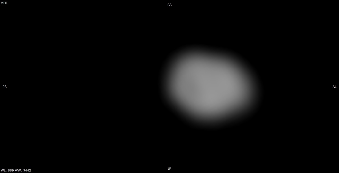
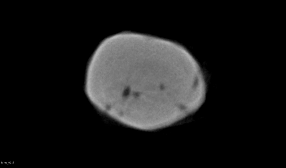
Fig. 1. Two-dimensional reconstructions of the apex of an extracted tooth in the axial section: a — CBCT: only one of the canals is visible; b — micro-CBCT: several microscopic canals
According to the CBCT data, inclusions were discovered inside the root canals in 14.70% of the cases (n=20). The results of micro-CBCT revealed inclusions in 72.79% of the roots (n=99). Some of the root canals contained dense microscopic elements. Additionally, amorphous low-density structures with occasional calcifications could be detected on some of the micro-CBCT images, which we interpreted as dental pulp inside of the root canals (Fig. 2).
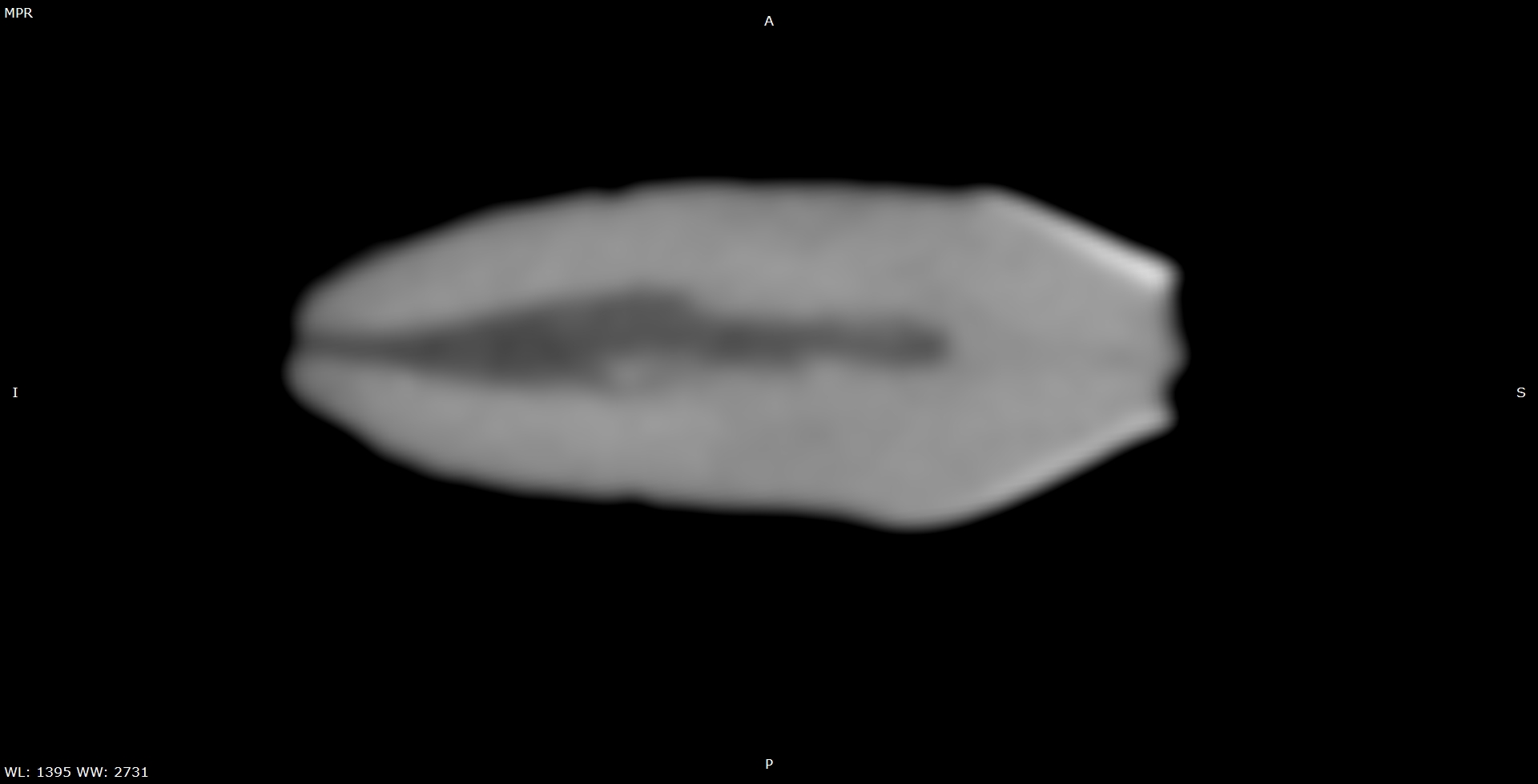
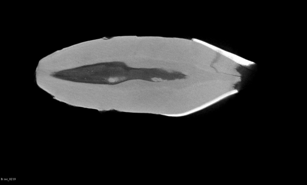
Fig. 2. Fragments of two-dimensional reconstructions of an extracted tooth in the longitudinal section: a — CBCT: the shades of gray inside the root canal are distributed unevenly, it is impossible to tell whether this should be interpreted as the contents of the canal or image noise; b — micro-CBCT: heterogeneous structure inside the root canal, its density is lower than dentin, with several high-density inclusions
The extracted teeth were subjected to physical force during removal and then preserved in the dried state, which resulted in many small and large dentinal cracks in the roots. Longitudinal fractures that reached root canals were found in 23.80% of the roots (n=29) on CBCT reconstructions and in 45.58% of the roots (n=62) on micro-CBCT images (Fig. 3).
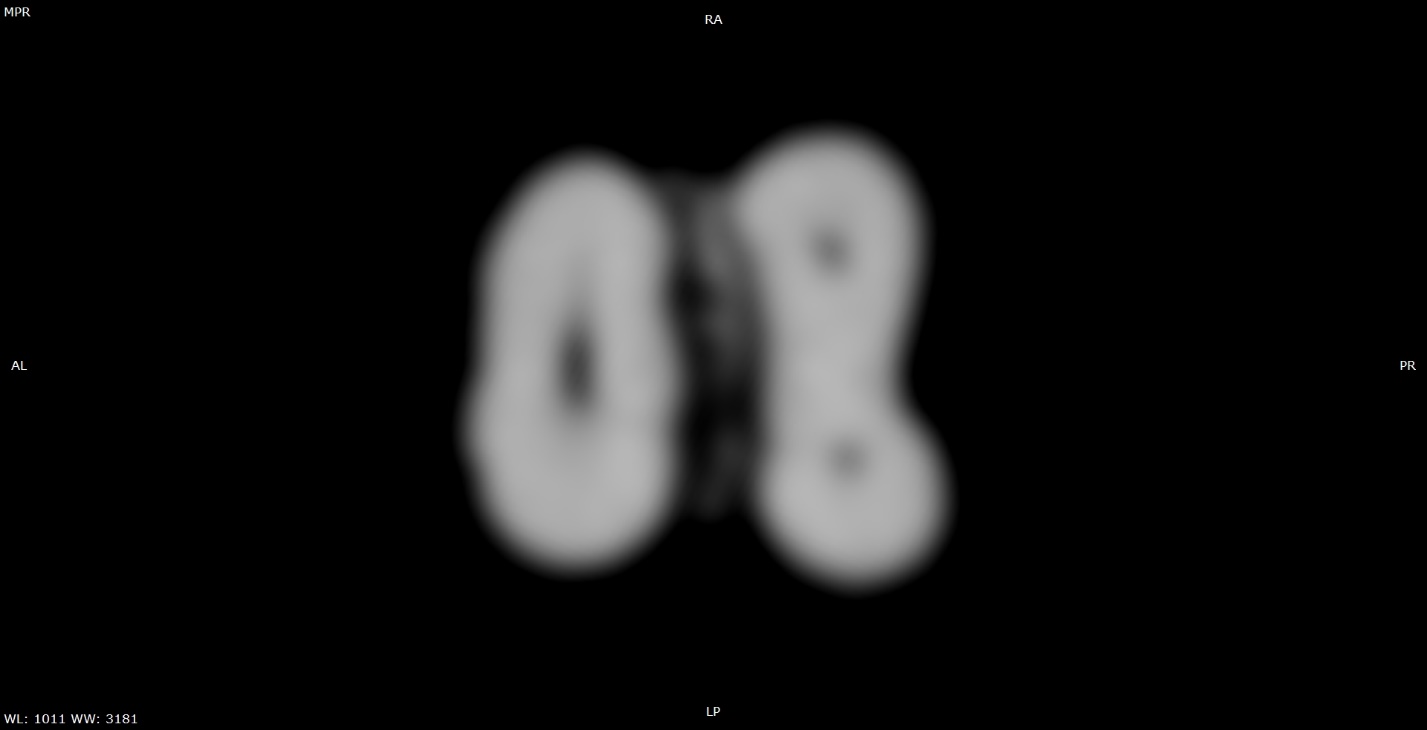
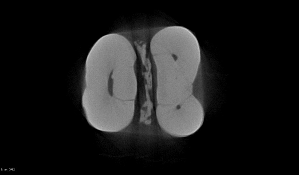
Fig. 3. Two-dimensional reconstructions of an extracted tooth in the axial section: a — CBCT: fracture line is practically indistinguishable; b — micro-CBCT: fracture line reaches the root canal
In the second stage of the study, multiplanar reconstructions were created based on CBCT and micro-CBCT scans of 91 endodontically treated roots. A description of radiological characteristics was summarized for each of the endodontic materials.
Calcium-aluminosilicate cement was homogeneous with blurred edges on CBCT images, but had clear outlines and granular structure with multiple high-density inclusions on micro-CBCT images (Fig. 4).
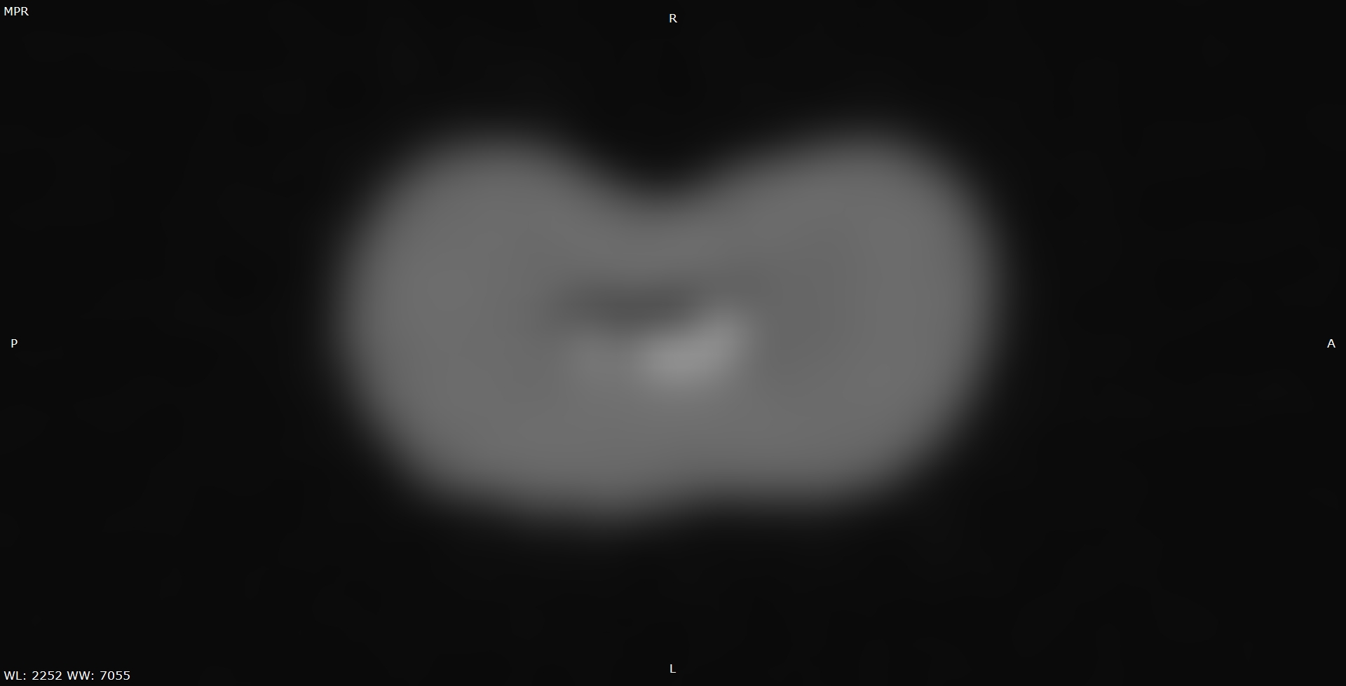
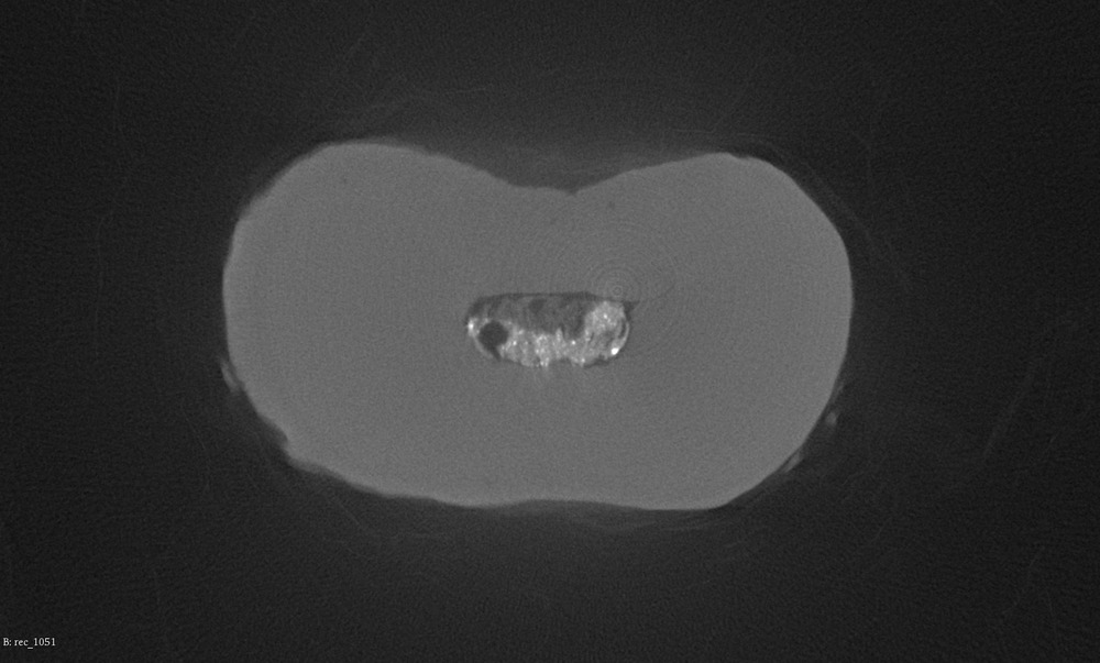
Fig. 4. Fragments of two-dimensional reconstructions of an extracted tooth in the axial section, the root canal is filled with calcium-aluminosilicate cement: a — CBCT: the filling material is homogeneous with blurred outlines, the root canal is partially filled with cement; b — micro-CBCT: granular structure of the filling material contains multiple high-density inclusions, as well as several voids of various shapes and sizes. The root canal is partially filled with dentin fragments that were not removed after mechanical preparation
Individual gutta-percha pins were not visible on cone beam computed tomograms of the roots obturated with cold lateral condensation of gutta-percha when several pins were inserted inside the same root canal. The zinc oxide-eugenol sealer was visually indistinguishable from gutta-percha. Micro-CBCT made separate gutta-percha pins visible regardless of their relative position and number, the pins merged only in tightly sealed canals, usually in the apical part. The sealer could be seen as a substance with a small number of high-density inclusions (Fig. 5 and 7).
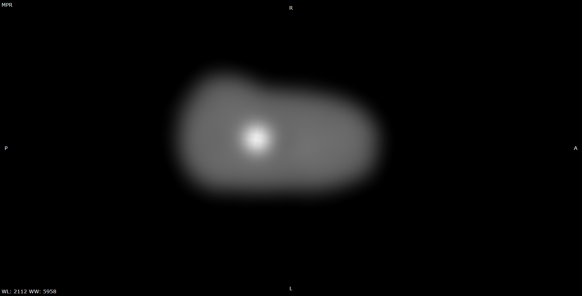
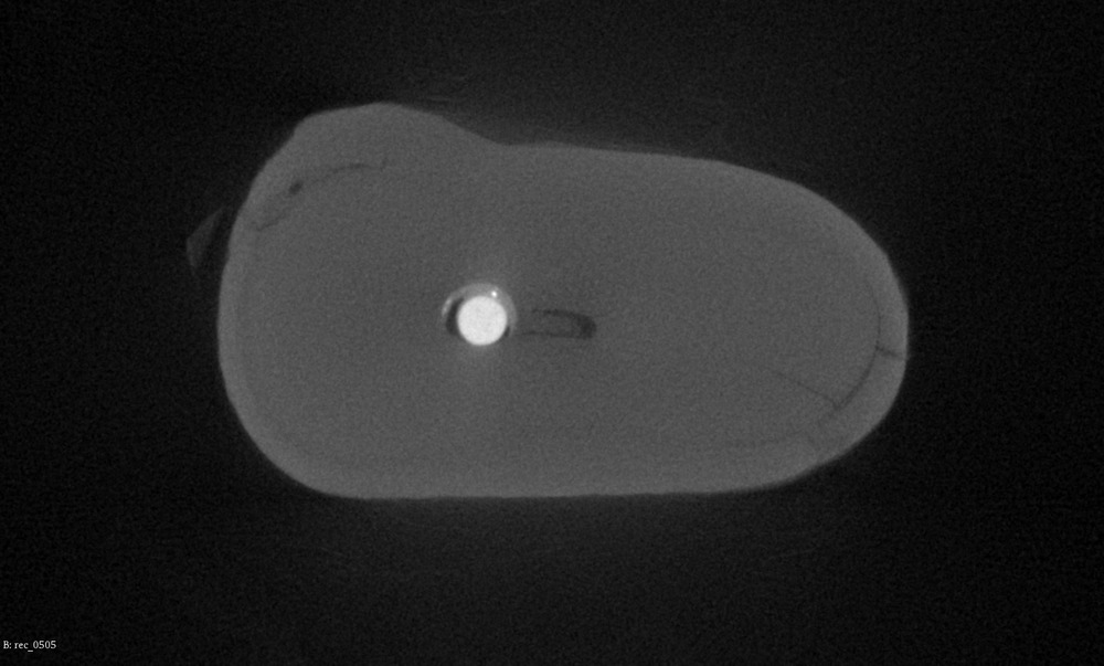
Fig. 5. Fragments of two-dimensional reconstructions of an extracted tooth in the axial section, the root canal is obturated with one gutta-percha pin for cold lateral condensation and a zinc oxide-eugenol sealer: a — CBCT: the endodontic materials are not visible, their outlines are blurred; b — micro-CBCT: a visible gap between the gutta-percha pin and the sealer. The sealer contains several microscopic hyperdense granules. The narrow root canal is partially filled with dentin fragments that were not removed after mechanical preparation
The microscopic details of the filling materials could not be visualized on CBCT images of the roots treated with thermoplasticized gutta-percha. The homogeneous content of the root canals caused moderate artifacts that blurred the image around the canals. Micro-CBCT images revealed the homogeneous substance of gutta-percha and dense plastic carriers. The outlines of the structures around the root canals were not distorted by artifacts. The epoxy-based sealer and thermoplasticized gutta-percha were visually identical except for their density, and the difference between gutta-percha and a thin layer of sealer was often impossible to determine on multiplanar reconstructions (Fig. 6).
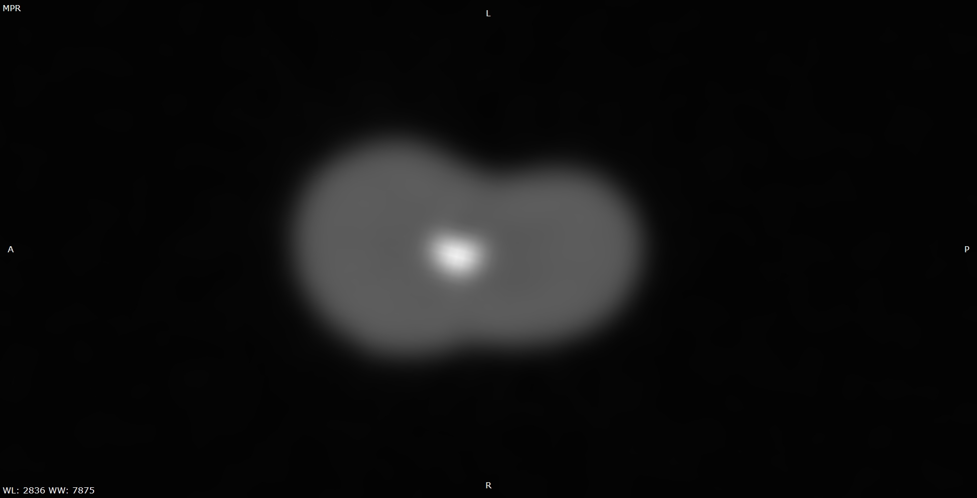
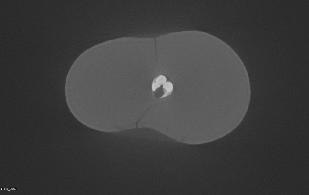
Fig. 6. Fragments of two-dimensional reconstructions of an extracted tooth in the axial section, the root canal contains thermoplasticized gutta-percha on a plastic carrier and an epoxy resin-based sealer: a — CBCT: the root canal is filled partially, the filling materials inside the root canal are blurred; b — homogenous gutta-percha is clearly divided from a plastic carrier, the root canal is partially filled, there is a layer of sealer between gutta-percha and dentin. The sealer spreads inside the fracture lines of the root. Gutta-percha forms a spiral shape because the obturator was rotated inside the canal
Voids inside the endodontic materials or between the filling materials and dentin were visible only on CBCT images of underfilled root canals. Micro-CBCT revealed microscopic voids inside and outside the endodontic materials even in fully and continuously filled root canals. Voids of various shapes and sizes were typically found inside the calcium-aluminosilicate cement and the zinc oxide-eugenol sealer (Fig. 4—6). Multiple small voids sometimes accumulated along the longitudinal axis of gutta-percha pins for cold lateral condensation, creating an appearance of hollow cores inside the pins. Some gutta-percha pins were torn in places where they bent and twisted, gutta-percha pins also retained their changed shape after being deformed by an external force (Fig. 7). Thermoplasticized gutta-percha contained rare microscopic voids and, in some cases, peeled off from its plastic carrier.
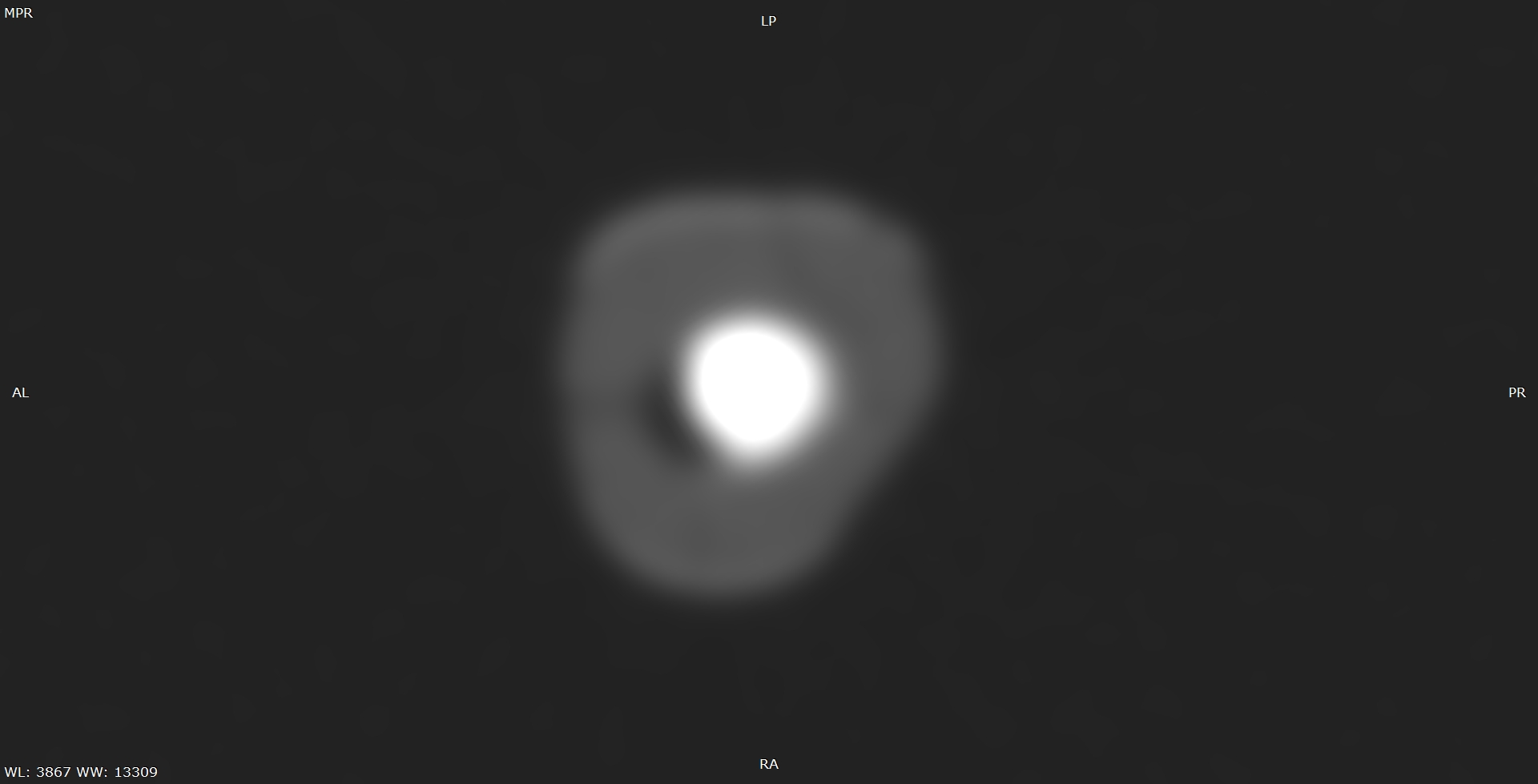
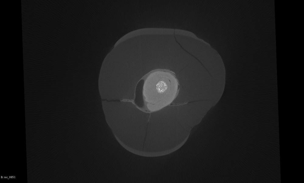
Fig. 7. Fragments of two-dimensional reconstructions of an extracted tooth in the axial section, the root canal contains gutta-percha cones for cold lateral condensation and a zinc oxide-eugenol sealer: a — CBCT: gutta-percha cones and the sealer are not visible, the outlines of the root canal are blurred; b — micro-CBCT: the sealer and the gutta-percha cones are clearly visible inside the root canal. The pins remain in a deformed state. Two of the pins appear to have hollow centers
Metal fragments were always round in shape on two dimensional CBCT images in the axial section, and their three-dimensional CBCT reconstructions had no surface detail. Metal artifacts blurred the details close to the broken instruments. Two-dimensional micro-CBCT images made it possible to visualize the rounded, triangular and tetragonal shapes of the instruments. Metal artifacts were noticeable, but only the small low-density structures near the surfaces of the metal fragments were hidden from view (Fig. 8 and Fig. 9).
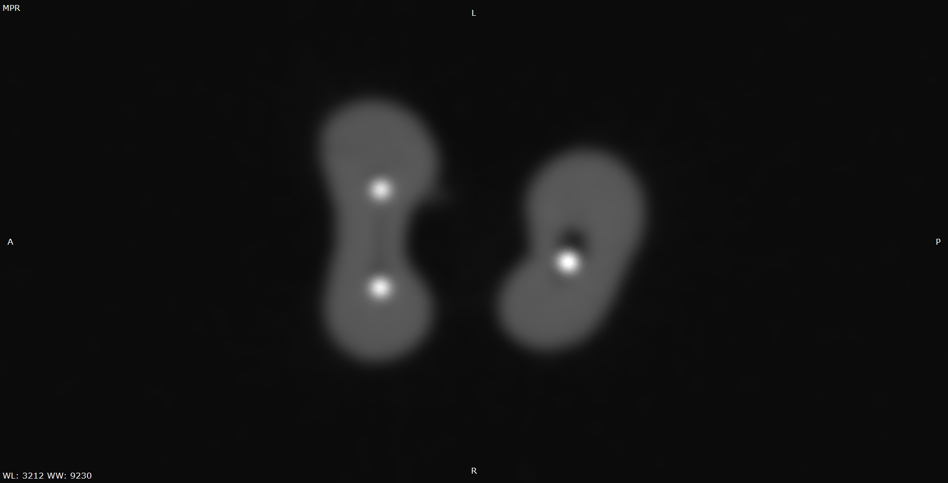
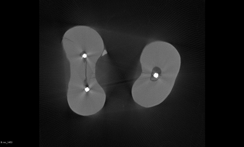
Fig. 8. Two-dimensional reconstructions of an extracted tooth in the axial section, the root canal contains a fragment of a metal instrument: a — CBCT: the metal fragment is round in the axial section; b — micro-CBCT: the metal fragment is tetragonal in the axial section, fragments of dentin inside the root canal and fracture lines in the root are clearly visible against the background of metal artifacts
It was impossible to determine the edges of the blurred outlines of metal fragments in CBCT images, which made building three-dimensional models of the broken instruments difficult. Visually it resulted in models with smooth featureless surfaces. Three-dimensional reconstructions created with the help of micro-CBCT were detailed and represented the shapes and sizes of metal fragments more accurately.
Three-dimensional models of metal fragments were built for 26 roots that contained broken instruments, and the volumes of the models were calculated in mm3 for CBCT and micro-CBCT. For each of the metal fragments, the volume of the CBCT model was always bigger. In 89.5% of the cases the volume of a model built with the use of CBCT was at least 3 times bigger than the volume calculated with the help of micro-CBCT (Fig. 9).
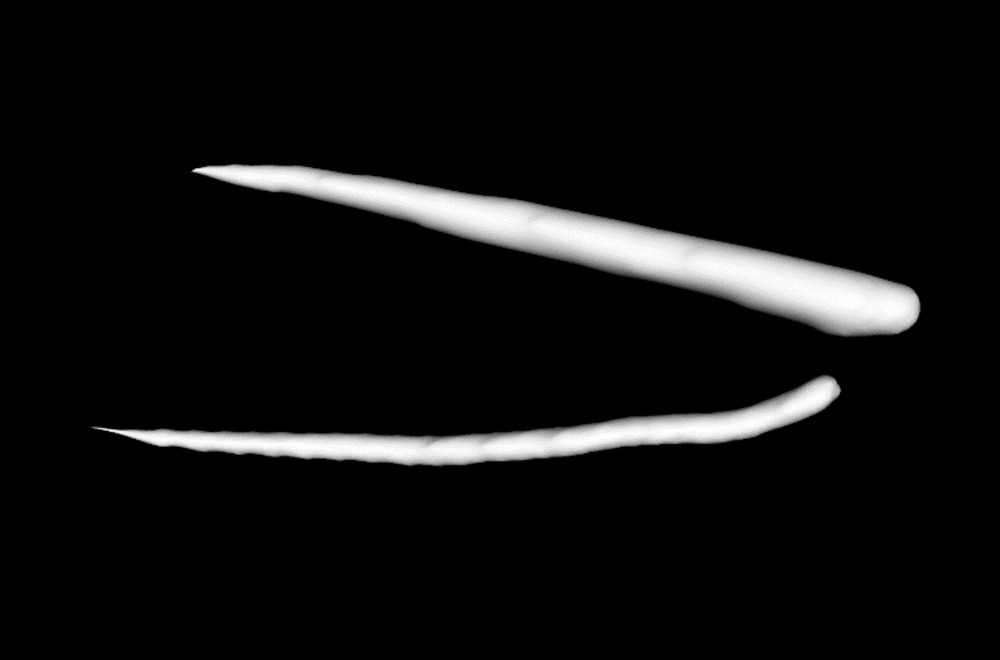
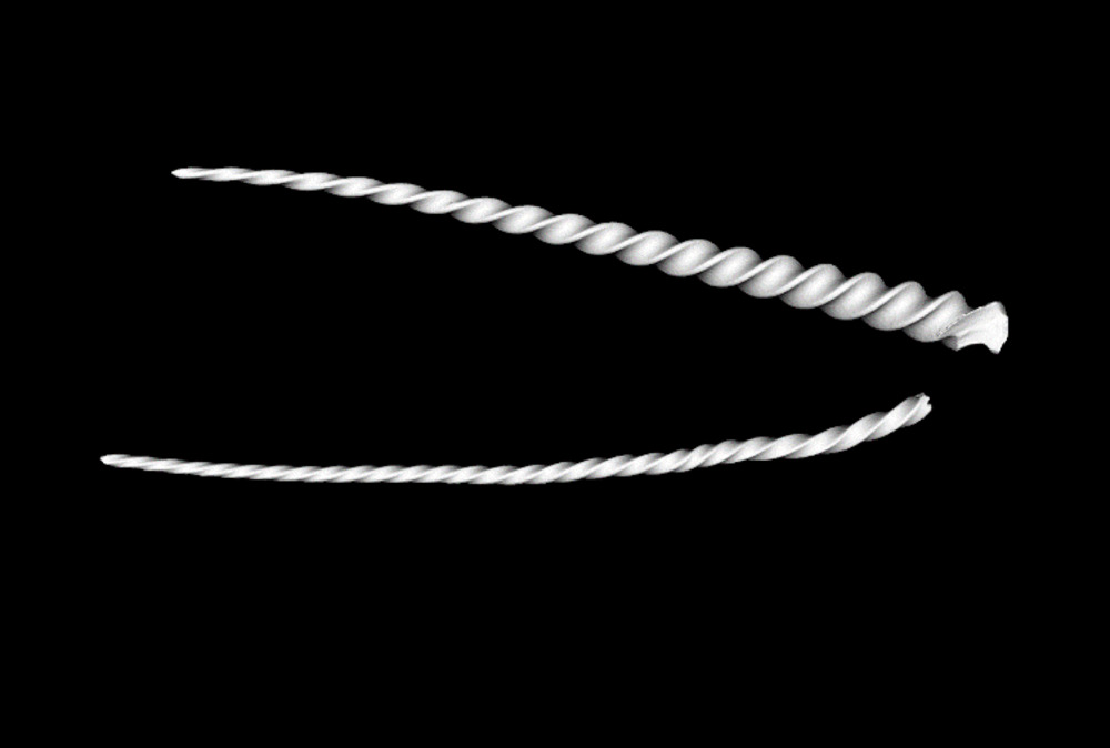
Fig. 9. Three-dimensional reconstruction of two broken metal instruments inside root canals of an extracted tooth: a — CBCT: the models show simplified shapes of the fragments, b — micro-CBCT: the fragments are spiral in shape, with visible details on the surface
Conclusion.
Micro-CBCT is characterized by a higher spatial resolution and better image clarity than CBCT. Micro-CBCT reveals additional root canals, fracture lines and inclusions inside root canals that are not visible on CBCT images.
CBCT allows to discover endodontically treated root canals and large filling defects, while micro-CBCT makes it possible to detect several filling materials inside one root canal and to characterize the microscopic structure of the materials.
Artifacts from hyperdense objects were less pronounced on micro-CBCT images compared to CBCT. Micro-CBCT data can be used to build detailed three-dimensional models of metal objects inside the root canals of extracted teeth.
References
- Vasilyev A.Yu., Petrovskaya V.V., Topolnitsky O.Z., Borovitskaya N.N. Cone-beam tomography of congenital fissures of the alveolar process and palate. — Journal of Radiology and Nuclear Medicine. — 2011; 2: 004—006 (In Russ.).
- Nechaeva N.K., Dolgalev A.A. Planning dental implantation on upper jaw by means of cone-beam tomography. — Medical alphabet. — 2018; 8 (345): 44—7 (In Russ.).
- Chogle S., Zuaitar M., Sarkis R., Saadoun M., Mecham A., Zhao Y. The Recommendation of Cone-beam Computed Tomography and Its Effect on Endodontic Diagnosis and Treatment Planning. — J Endod. — 2020; 46 (2): 162—8. PMID: 31837812
- Patel S., Brown J., Pimentel T., Kelly R.D., Abella F., Durack C. Cone beam computed tomography in Endodontics — a review of the literature. — Int Endod J. — 2019; 52 (8): 1138—52. PMID: 30868610
- Yeung A.W.K., Jacobs R., Bornstein M.M. Novel low-dose protocols using cone beam computed tomography in dental medicine: a review focusing on indications, limitations, and future possibilities. — Clin Oral Investig. — 2019; 23 (6): 2573—81. PMID: 31025192
- Dolgalev A.A., Nechaeva N.K., Ivancheva E.N., Nagoryansky V.Yu. The use of cone beam computed tomography in endodontics (Part I). Analysis of root canal topography. — Endodontics Today. — 2017; 1: 68—71 (In Russ.).
- Dolgalev A.A., Nechaeva N.K., Ivancheva E.N. The use of cone beam computed tomography in endodontics (Part II). Analysis of root canal topography. — Endodontics Today. — 2017; 2: 69—73 (In Russ.).
- Solovyova O.A., Vinnichenko Yu.A., Goman M.V., Dolgalev A.A., Zaborovets I.A. The use of cone beam computed tomography in endodontics (Part III). The instrumental method of treatment of the canal if it contains fragments tool. — Endodontics Today. — 2017; 3: 29—33 (In Russ.).
- Obodovskij A.V. Development and research of technical means of microfocus x-ray tomography: PhD dissertation. — Saint-Petersburg, 2018. — 135 p. (In Russ.).
- Bayram H.M., Bayram E., Ocak M., Uzuner M.B., Geneci F., Celik H.H. Micro-computed tomographic evaluation of dentinal microcrack formation after using new heat-treated nickel-titanium systems. — J Endod. — 2017; 43 (10): 1736—9. PMID: 28756963
- Castagnola R., Marigo L., Pecci R., Bedini R., Cordaro M., Coppola E.L., Lajolo C. Micro-CT evaluation of two different root canal filling techniques. — Eur Rev Med Pharmacol Sci. — 2018; 22 (15): 4778—83. PMID: 30070311
- Elenjikal M.J., Latheef A.A., Kader M.A.M., Ganapathy S., Mohamed A.B., Sainudeen S.S., Abdulla A.M., Saquib S.S. A comparative evaluation of five obturation techniques in the management of simulated internal resorptive cavities: An ex vivo study. — J Pharm Bioallied Sci. — 2019; 11 (Suppl 2): S450—6. PMID: 31198386
- Irie M.S., Rabelo G.D., Spin-Neto R., Dechichi P., Borges J.S., Soares P.B.F. Use of micro-computed tomography for bone evaluation in dentistry. — Braz Dent J. — 2018; 29 (3): 227—38. PMID: 29972447
- Jho W., Park J.-W., Kim E., Song M., Seo D.-G., Yang D.-K., Shin S.-J. Comparison of root canal filling quality by mineral trioxide aggregate and gutta percha cones/AH plus sealer. — Dent Mater J. — 2016; 35 (4): 644—50. PMID: 27477231
- Kierklo A., Tabor Z., Pawińska M., Jaworska M. A microcomputed tomography-based comparison of root canal filling quality following different instrumentation and obturation techniques. — Med Princ Pract. — 2015; 24 (1): 84—91. PMID: 25359228
- Lacerda M.F.L.S., Marceliano-Alves M.F., Pérez A.R., Provenzano J.C., Neves M.A.S., Pires F.R., Gonçalves L.S., Rôças I.N., Jr J.F.S. Cleaning and shaping oval canals with 3 instrumentation systems: A correlative micro-computed tomographic and histologic study. — J Endod. — 2017; 43 (11): 1878—84. PMID: 28951035
- Leoni G.B., Versiani M.A., Silva-Sousa Y.T., Bruniera J.F.B., Pécora J.D., Sousa-Neto M.D. Ex vivo evaluation of four final irrigation protocols on the removal of hard-tissue debris from the mesial root canal system of mandibular first molars. — Int Endod J. — 2017; 50 (4): 398—406. PMID: 26992452
- Meng Y., Xu J., Pradhan B., Tan B.K., Huang D., Gao Y., Zhou X. Microcomputed tomographic investigation of the trepan bur/microtube technique for the removal of fractured instruments from root canals without a dental operating microscope. — Clin Oral Investig. — 2020; 24 (5): 1717—25. PMID: 31346785
- Neves A.B., Bergstrom T.G., Fonseca-Gonçalves A., Dos Santos T.M.P., Lopes R.T., de Almeida Neves A. Mineral density changes in bovine carious dentin after treatment with bioactive dental cements: a comparative micro-CT study. — Clin Oral Investig. — 2019; 23 (4): 1865—70. PMID: 30218229
- Jr J.F.S., Pérez A.R., Marceliano-Alves M.F., Provenzano J.C., Silva S.G., Pires F.R., Vieira G.C.S., Rôças I.N., Alves F.R.F. What happens to unprepared root canal walls: a correlative analysis using micro-computed tomography and histology/scanning electron microscopy. — Int Endod J. — 2018; 51 (5): 501—8. PMID: 28196289
- Rossi-Fedele G., Ahmed H.M.A. Assessment of root canal filling removal effectiveness using micro-computed tomography: A systematic review. — J Endod. — 2017; 43 (4): 520—6. PMID: 28214018
- Suguro H., Takeichi O., Hayashi M., Okamura T., Hira A., Hirano Y., Ogiso B. Microcomputed tomographic evaluation of techniques for warm gutta-percha obturation. — J Oral Sci. — 2018; 60 (2): 165—169. PMID: 29657249
- Torres F.F.E., Bosso-Martelo R., Espir C.G., Cirelli J.A., Guerreiro-Tanomaru J.M., Tanomaru-Filho M. Evaluation of physicochemical properties of root-end filling materials using conventional and Micro-CT tests. — J Appl Oral Sci. — 2017; 25 (4): 374—80. PMID: 28877275
- de Faria Vasconcelos K., Dos Santos Corpas L., da Silveira B.M., Laperre K., Padovan L.E., Jacobs R., de Freitas P.H.L., Lambrichts I., Bóscolo F.N. MicroCT assessment of bone microarchitecture in implant sites reconstructed with autogenous and xenogenous grafts: a pilot study. — Clin Oral Implants Res. — 2017; 28 (3): 308—13. PMID: 26932194
- Velozo C., Albuquerque D. Microcomputed tomography studies of the effectiveness of XP-endo shaper in root canal preparation: A review of the literature. — ScientificWorldJournal. — 2019; 2019: 3570870. PMID: 31531000
- Zuolo M.L., De-Deus G., Belladonna F.G., da Silva E.J.N.L., Lopes R.T., Souza E.M., Versiani M.A., Zaia A.A. Micro-computed tomography assessment of dentinal micro-cracks after root canal preparation with TRUShape and self-adjusting file systems. — J Endod. — 2017; 43 (4): 619—22. PMID: 28216274
- Domenyuk D.A., Chukov S.Z., Anfinogenova O.I., Rzhepakovsky I.V., Ivanyuta O.O. Application of computer microtomography in the study of morphostructural peculiarities of hard tissues of teeth in early forms of carious lesions. — Kuban Scientific Medical Bulletin. — 2018; 6 (25): 57—67 (In Russ.).
- Domenyuk D.A., Davydov B.N. Possibilities of microcomputer tomography in the diagnostics of early forms of caries of a chewing surface of permanent molars in children. Part I. — Pediatric Dentistry and Profilaxis. — 2018; 4 (67): 61—4 (In Russ.).
- Domenyuk D.A., Davydov B.N. Possibilities of microcomputer tomography in the diagnostics of early forms of caries of a chewing surface of permanent molars in children. Part II. — Pediatric Dentistry and Profilaxis. — 2019; 2 (70): 4—12 (In Russ.).
- Vasilev A.Yu., Petrovskaya V.V., Khizhnyak A.Yu., Silyagina A.S., Potrakhov N.N. Analysis of endodontic treatment of teeth using various methods of radiology (in the experiment). — Biotechnosfera. — 2017; 5 (53): 57—61 (In Russ.).
- Vasilyev A.Yu., Petrovskaya V.V. Micro-CT as a new promising technology in dentistry. — Diagnostic Radiology and Radiotherapy. — 2018; 1 (9): 62—3 (In Russ.).
- Levitskaya A.D., Syutkina E.S., Gileva O.S., Galkin S.V., Efimov A.A., Savitskiy Ya.V. The evaluation of microstructure and mineral density of the focus of artificial enamel caries using X-ray computer microtomography. — Russian Journal of Biomechanics. — 2018; 4 (22): 485—502 (In Russ.).
- Mitronin A.V., Sobkina N.A., Pomeshcshikova N.I., Dmitrieva L.A. Use of computer microtomography to assess the quality of endodontic tooth treatment using modern instruments. — Endodontics Today. — 2018; 1: 22—6 (In Russ.).
- Petrovskaya V.V., Potrakhov N.N., Vasil’ev A.Yu. Cone beam computed tomography in the analysis of endodontic treatment of teeth (in an experiment). — Journal of Radiology and Nuclear Medicine. — 2019; 2 (100): 89—94 (In Russ.).
- Sobkina N.A., Pomeschikova N.I., Dmitrieva L.A. Analysis of root canal preparation using the ProTaper instruments. — Russian Stomatology. — 2018; 3 (11): 49—52 (In Russ.).


