
DOI:
10.37988/1811-153X_2020_4_43Dynamics of changes in the level of pain in the masticatory muscles in patients with reduced interalveolar height
Abstract
The aim of the study was to investigate the dynamics of pain intensity changes by a visual- analog scale (VAS) in patients with reduced vertical dimension of occlusion, who were treated using elastic bands and splints. Materials and methods. 52 people (15 men and 37 women) with reduced vertical dimension of occlusion and masticatory muscle hypertension were examined. The diagnosis was made on the basis of clinical and instrumental (electromyography) methods. Pain intensity was determined using VAS. The control group consist of 31 practically healthy people aged 16 to 31 years. Preliminary treatment of patients of the main group was carried out using elastic bands and splints. Results. It was found that the proposed method of masticatory muscle hypertension treatment in patients with reduced vertical dimension of occlusion provides reliable (Z1—3= –6.40; p1—3=0.01) reducing of pain intensity, tension and fatigue of the masticatory muscles by the end of second week of therapy (from 8.17±0.6 to 4.54±0.5 points by the VAS scale). It is also confirmed by reliable decrease of the bioelectric activity of the masticatory muscles. Conclusion. Thus, the proposed method of treatment of masticatory muscle hypertension in patients with reduced vertical dimension of occlusion has features, favorably distinguish it from the other methods of the therapy, because it provides gentle functioning condition for masticatory muscles.
Key words:
reduced vertical dimension of occlusion, masticatory muscle hypertension, visualanalog scale (VAS), elastic band, splintFor Citation
Introduction
Practicing dentists know that the treatment of masticatory muscle hypertension in patients with reduced interalveolar height is particularly difficult [1—4]. Even complex therapy with the involvement of various specialists does not provide an absolute result. A new promising direction for the treatment of these diseases may be the use of elastic bands [5—7].
The purpose of the research: to study the dynamics of changes in the intensity of pain in the masticatory muscles on the VAS scale in the treatment of patients with reduced interalveolar height using elastic bands and splints.
Materials and methods
There were examined 52 people (15 men and 37 women) aged from 21 to 65 years. The diagnosis was made based on the results of clinical and paraclinical (electromyography) research methods. Pain intensity was assessed using a visual-analog scale (VAS). The control group consisted of 31 practically healthy people aged 16 to 31 years.
The visual-analog pain scale is a subjective method of assessing the severity of pain syndrome, which has high validity and reliability [8]. The scale is a continuous segment of 10 cm in length, the initial mark of which corresponds to the absence of pain, and the end point reflects excruciating, unbearable pain. The patient was asked to put a mark on the scale, which, in his opinion, corresponds to the intensity of pain. Then, using a ruler, the distance (in mm) from the starting point to the obtained mark was measured. Several approaches have been proposed to interpret the results. We used the classification of Jensen et al. [9]: 0—4 mm — no pain; 5—44 mm — mild pain; 45—74 mm — moderate pain; 75—100 mm — severe pain. At the same time, according to the digital evaluation scale, 1—4 points correspond to mild pain, 5—6 points — moderate, 7—10 points — severe.
The criteria for inclusion in the research were: patients with reduced interalveolar height, suffering from pain, tension, fatigue, hypertension of the masticatory muscles lasting at least 1 month.
The criteria for non-inclusion were: a history of TMJ surgery (arthroscopy, arthrocentesis), TMJ trauma, systemic inflammatory diseases of the TMJ (rheumatoid arthritis), patients who had already been treated for parafunctions of the masticatory muscles in the last 6 months, inflammatory skin diseases at the site of the intended application of elastic bands, allergic reactions to acrylic adhesive gel, which is part of the tapes, individual intolerance to elastic bands.
To check the normality of the distribution, the Kolmogorov-Smirnov criterion was used. The nonparametric Mann-Whitney test was used to analyze the differences between the pain intensity levels at the treatment stages and to assess changes in the bioelectric activity of the masticatory muscles.
The main complaint in the studied patients was pain in the masticatory muscles, their rapid fatigue, tension, limited opening of the mouth.
During external examination, 38 (73.1%) patients had an increased degree of development of masticatory muscles (hypertrophy), their volume. In 25 people (48.1%), a reduction of m. masseter was observed periodically under the skin. In 32 (61.5%) subjects, there was a significant facial asymmetry associated with a shift of the chin to the right or left, as well as due to the unequal degree of development of the right and left masticatory muscles. The majority of patients (37 people, 71.1%) had a decrease in the lower part of the face, deepening of the nasolabial and chin folds, and drooping corners of the mouth. At the initial examination, the height of functional rest of the masticatory muscles was not determined in 41 people (78.8%). Limited mouth opening (less than 40 mm) was detected in 29 (55.7%) patients.
Palpation of the masticatory and lateral pterygoid muscles revealed their increased tension (47 people, 90.3%), while in some areas of the muscle, or rather, points — there was severe pain (“trigger” zones — areas where pressure causes spasm and pain).
It should be noted that 33 (63.4%) of the subjects were found to have teeth imprints on the lateral surface of the tongue and the mucous membrane of the cheeks. Generalized form of various degrees of increased erasability of dentition was observed in 45 (88.6%) patients, localized — in 7 (13.4%) patients. Wedge-shaped defects were observed in 27 (51.9%) patients.
Relief of masticatory muscle hypertension was decided to be carried out using elastic bands and splints. We have developed a technique for applying tapes and a Protocol for their use. Kappa was used by patients constantly (with the exception of food intake) throughout the duration of treatment.
Method of fixing elastic bands
We used Kinexib Ultraviolet elastic bands (Suzhou Sunmed, China) approved by Russian Federal Service for Surveillance in Healthcare (registration certificate for medical device No. RZN 2019/8334 dated April 26, 2019). The width of the elastic bands is 5 cm; the roll of tapes has a breakaway line every 25 cm. According to the manufacturer, the elastic bands consist of 97% viscose and 3% cotton; one of the sides is coated with hypoallergenic acrylic glue, which is activated at body temperature.
The elastic band is similar in thickness and degree of stretching to the epidermis. It does not contain medicinal substances and is water-resistant. High-quality elastic tape stretches only in one direction — along the longitudinal axis. The application of the tapes lasts up to 5 days, after which the elastopolymer that is part of it loses its elasticity. The elastic band provides a therapeutic effect 24 hours a day throughout the entire time of use [10].
Elastic tape can be applied with a tension of 0 to 100%, however, the maximum degree of tension (76—100%) is rarely used. When applying tapes to the TMJ and masticatory muscles, the degree of tension can vary depending on the tone of the masticatory muscles, General physical fitness, weight and body volume of the patient.
We used Y-shaped tapes. In this case, the upper strip of tape should cover the point of the beginning of the masticatory muscles (zygomatic process of maxilla and zygomatic arch) and the lower point of its end (the angle of the mandible). The use of other types of tapes (for example, I-shaped) does not allow to fully cover both points of attachment of the masticatory muscle itself.
The distance between the upper and lower bands of the Y-shaped tape fixed on the face of patients is individual for each of them and depends on the type of structure of the face, its size, and shape (Fig. 1).
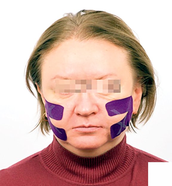 a
a
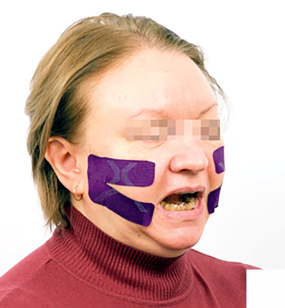 b
b
Each application of elastic bands lasted for three days for 6 weeks with a one-day break every two procedures. Patients were trained in the technique of applying tapes and provided with a memo developed by us for self-execution of applications.
The effectiveness of treatment was evaluated on days 1, 7, 14, 21 and 42 by analyzing the dynamics of patient complaints, pain intensity using VAS and bioelectric activity of the masticatory muscles.
Results
Patients were surveyed using VAS every 7 days for 42 days.
At the beginning of treatment, the level of pain in the masticatory muscles was 8.17±0.6 points on the VAS scale, which corresponds to its strong intensity. Patients during this period are characterized by the following symptoms: limited opening of the mouth (15—17 mm between the cutting edges of the incisors of the upper and lower jaws); blocking of movements of the lower jaw; aching prolonged pain that occurs both during movements of the lower jaw and at rest; “trigger” zones detected by palpation of the masticatory muscles; soreness, rapid fatigue, tension of the masticatory muscles, expressed more often in the morning, after sleep; sensation of individual teeth, pain when biting on some teeth; decreased performance, headache, sleep disturbance.
It should be noted that the external examination of patients showed a decrease in the height of the lower face, while the height of the functional rest of the masticatory muscles was not determined in 41 of 52 people (78.8%).
By the end of the first week, on day 7, VAS indicators were 7.27±0.5 points (the upper limit of the interval corresponding to moderate pain intensity). The clinical symptoms remained the same.
The tipping point of treatment (V1—3= −6.40; p1—3=0.01), aimed at reducing the level of pain, was determined by the end of the second week (on day 14) of treatment and was 4.54±0.5 points on the VAS scale (the interval corresponding to the lower limit of moderate pain intensity). By this time, 37 out of 52 patients (71.1%) of the main group had a marked decrease in the severity of pain in the masticatory muscles. It became possible to determine the functional height of the face in 31 of 41 (75.6%) subjects. At this stage of treatment, the clinical picture was the following: the opening of the mouth increased to 28—30 mm; palpation of the masticatory muscles and TMJ became moderately painful; aching pain in the masticatory muscles occurred mainly during the movements of the lower jaw; the frequency of headaches decreased, sleep normalized, and performance increased.
On day 21 of treatment, the level of pain intensity was 3.27±0.6 points (the interval corresponds to a weak pain severity). Characteristic clinical signs were as follows: increased mouth opening to 31—36 mm; palpation of the masticatory muscles and TMJ became almost painless; pain in the masticatory muscles and TMJ occurred rarely and mainly against the background of physical and emotional stress; palpation of the masticatory muscles and TMJ became painless; headaches practically did not occur.
On day 42 of the study, a significant reduction in pain in the masticatory muscles was recorded: it was 2.12±0.5 points (V1—5= −6.46; p1—5=0.01; interval of mild pain severity). The opening of the mouth in patients increased to 42—45 mm, pain in the masticatory muscles and TMJ was absent (or extremely rare), palpation of the masticatory muscles became painless, and headaches did not occur (Fig. 2).

After treatment with elastic bands and splint, the indicators of the maximum amplitude of bioelectric activity of the masticatory muscles had a significant decrease in comparison with the initial values (see table).
| Masticatory muscles | Group of patients |
V2—3 p2—3 |
|||
|
Control (n=31) |
Main before treatment (n=52) |
Main, Day 14 of treatment (n=52) |
|||
| 1 | 2 | 3 | |||
| Masseter | right | 5.7±0.07 | 11.4±0.19 | 5.7±0.14 |
−6.30 0.015 |
| left | 5.5±0.13 | 10.2±0.18 | 5.8±0.21 |
−6.29 0.01 |
|
| Temporals | right | 4.2±0.11 | 10.5±0.15 | 4.1±0.19 |
−6.39 0.01 |
| left | 4.1±0.09 | 9.9±0.16 | 4.3±0.18 |
−6.28 0.015 |
|
Discussion
Evaluation of the result of treatment of masticatory muscle hypertension of patients with reduced interalveolar height using elastic bands and splints showed a positive effect of therapy.
The use of elastic bands has proven to be a simple and effective method of local action on spasmodic muscles, as well as in intermittent muscle hypertension.
According to the literature, the therapeutic effect of elastic bands is based on the following mechanisms:
- activation of microcirculation in the skin, underlying connective tissue and intercellular substance, elimination of metabolic products, improvement of lymphatic drainage due to the elastic properties of the tapes due to the lifting effect;
- reduction of pain syndrome (according to the theory of portal pain control by R. Melzack and P. Wall, 1965);
- restoration of functional activity of muscles and normalization of joint function by regulating the afferent flow coming from their proprioreceptors [7, 11].
We suppose that, apart from the mechanisms of action of elastic bands described in the literature, another one is realized: they support the muscles, thus reducing their tone, as a result of which the provocation to pain is sharply reduced or disappears altogether. With hypertension of the masticatory muscles, the main thing is to create a gentle regime for its moderate work. Gentle mode — a condition in which an elastic band reduces the functional overload of the muscle, forcing it to work as in a healthy person [12].
Applications of elastic bands can be recommended for use at the preventive stage of treatment of hypertension of the masticatory muscles, since, despite the possibility of reducing the symptoms of hypertension, the risk of their recurrence remains with repeated mental injuries, chronic emotional stress.
Conclusion
Using elastic bands and splints in the treatment of masticatory muscle hypertension in patients with reduced interalveolar height has proven to be a rather effective treatment method. Among the main results of treatment, the following can be marked: 1) relieving pain and hypertension of the masticatory muscles; 2) achieving their relaxation; 3) normalizing the height of the lower part of the face; 4) accelerating the rehabilitation process.
References
- Voityatzkaya I.V., Tsimbalistov A.V. Reduced occlusion syndrom. — Head Neck. — 2017; 3: 46—50 (In Russ.).
- Bulycheva E.A., Chikunov S.O., Alpateva Yu.V. Rehabilitation therapy development in patients with temporomandibular joint dysfunction, complicated by masticatory muscle hypertension (III part). — The Dental Institute. — 2013; 2: 44—5 (In Russ.).
- Alpateva Yu.V. Rehabilitation of the patients with reduced vertical dimensuon of occlusion: Synopsis of a master’s thesis. — Moscow.: Moscow State University of Medicine and Dentistry, 2017. — 23 p. (In Russ.).
- Orlova O.R., Soykher M.I., Soykher M.G., Mingazova L.R., Medovnikova D.V., Soykher E.M., Konovalova Z.N., Alekseeva A.Yu. Bruxism: methods of application and results of treatment with botulinum neuroprotein (relatox). — Neuromuscular diseases. — 2019; 9 (2): 12—20. (In Russ.).
- Iordanishivili A.K., Serikov A.A. The kinesiotaping in periodontics and dental artrologie. — Parodontology. — 2018; 24 (3): 80—83 (In Russ.).
- Bae Y. Change the myofascial pain and range of motion of the temporomandibular joint following kinesio taping of latent myofascial trigger points in the sternocleidomastoid muscle. — J Phys Ther Sci — 2014; 26 (9): 1321—4. DOI: 10.1589/jpts.26.1321
- Wu W.-T., Hong C.-Z., Chou L.-W. The Kinesio Taping Method for Myofascial Pain Control. — Evidence-Based Complementary and Alternative Medicine. — 2015; 2015: 950519. DOI: 10.1155/2015/950519
- Boonstra A.M., Preuper H.R.S., Reneman M.F., Posthumus J.B., Stewart R.E. Reliability and validity of the visual analogue scale for disability in patients with chronic musculoskeletal pain. — Int J Rehabil Res. — 2008; 31 (2): 165—9. DOI: 10.1097/MRR.0b013e3282fc0f93
- Jensen M.P., Chen C., Brugger A.M. Interpretation of visual analog scale ratings and change scores: a reanalysis of two clinical trials of postoperative pain. — J. Pain. — 2003; 4 (7): 407—14. DOI: 10.1016/s1526-5900(03)00716-8
- Shpekht M.V., Pirogova L.A. Methodological aspects of combined use of rehabilitation measures in the recovery of postoperative menisci. — J Grodno State Med University. — 2020; 18 (2): 137—46 (In Russ.).
- Williams S., Whatman C., Hume P.A. et al. Kinesio taping in treatment and prevention of sports injuries: a meta-analysis of the evidence for its effectiveness. — Sports Med. — 2012, 42: 153—64. DOI: 10.2165/11594960-000000000-00000.
- Trezubov V.N., Bulycheva E.A., Bystrova Yu.A., Gorbachev V.V. Biofeedback therapy in patients with temporomandibular joint dysfunction. — The Dental Institute. — 2003; 3: 31—5. (In Russ.).


